

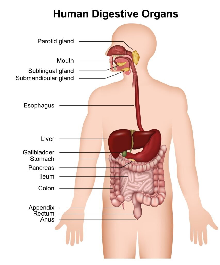
The human digestive system is a complex network of organs working together to break down the food we eat into absorbable molecules. This process, known as digestion, involves both mechanical and chemical actions. It all begins in the mouth, where teeth mechanically chew food into smaller pieces, while the salivary glands secrete saliva. Saliva contains an enzyme called salivary amylase which initiates the chemical digestion of starch into simpler sugars. The tongue then rolls the food into a soft, wet ball called a bolus, pushing it to the back of the throat to be swallowed into the esophagus. The esophagus doesn’t produce any digestive enzymes but uses wave-like muscular contractions, known as peristalsis, to transport the bolus down into the stomach.
The stomach acts as a muscular, J-shaped bag that serves as a mixing and churning chamber. Its inner lining secretes gastric juice, which includes hydrochloric acid, the enzyme pepsin, and mucus. The acid creates an acidic environment that kills most harmful bacteria and activates pepsin. Pepsin is crucial as it begins the digestion of proteins, breaking them down into peptides. The stomach’s muscular walls churn the food, mixing it thoroughly with the gastric juices to form a semi-liquid, acidic substance called chyme. This chyme is then released in small amounts through the pyloric sphincter into the next part of the digestive tract, the small intestine.
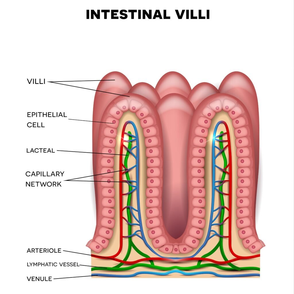
The small intestine is the primary site for both the complete digestion of food and the absorption of nutrients. It receives secretions from two major organs: the liver, which produces bile that emulsifies fats, and the pancreas, which sends pancreatic juice containing a variety of enzymes to break down starch, proteins, and fats. The inner wall of the small intestine is lined with millions of tiny, finger-like projections called villi, which dramatically increase the surface area for absorption. Here, the final products of digestion—like glucose, amino acids, and fatty acids—are absorbed into the bloodstream. The remaining undigested and unabsorbed material then moves into the large intestine, where water and some salts are reabsorbed, before the waste is temporarily stored in the rectum and expelled through the anus.
Multiple choice type
Question 1.
Pylorus is an opening from
- esophagus into stomach
- mouth cavity into stomach
- stomach into intestine
- intestine into rectum
Question 2.
Gastric juice contains
- HCl and pepsin
- pepsin and trypsin
- trypsin and HCl
- amylopsin and pepsin
Question 3.
The water from the digested food is mainly absorbed by
- stomach
- duodenum
- colon
- Rectum
Question 4.
Which one of the following pairs of types of teeth perform one common function as stated against it?
- Incisors, canines – Holding
- Canines, premolars – Biting
- Premolars, molars – Grinding
- Molars, incisors – Tearing
Very short answer type
Question 1.
What is the dental formula of a normal human adult?
Ans:
The dental formula of a normal human adult is 2123 / 2123.
Explanation
This formula represents the number of teeth in one half of the upper jaw (numerator) and one half of the lower jaw (denominator). The digits correspond to the four types of teeth, moving from the center outwards:
- 2 = Incisors (I)
- 1 = Canine (C)
- 2 = Premolars (P)
- 3 = Molars (M)
Multiplying this count (8 teeth per half jaw) by four quadrants gives a total of 32 permanent teeth.
Question 2.
Mention two reflexes which occur when a person chews and swallows food.
Ans:
The process of chewing and swallowing involves a complex sequence of coordinated movements, including both voluntary actions (like initial chewing) and involuntary reflexes.
Here are two key reflexes that occur when a person chews and swallows food:
1. Salivary Reflex (Chewing)
- Stimulus: The presence of food (taste, smell, sight, or physical presence in the mouth) or the act of chewing.
- Action: This reflex triggers the rapid and increased secretion of saliva from the salivary glands (parotid, submandibular, sublingual).
- Function: Saliva moistens the food, making it easier to chew and swallow, and begins the chemical digestion of starches via the enzyme amylase.
2. Pharyngeal (Swallowing) Reflex (Swallowing)
- Stimulus: The presence of a food bolus (chewed mass of food) hitting the back of the pharynx (throat).
- Action: This reflex, which is entirely involuntary, coordinates several rapid movements to ensure food goes down the esophagus and not the trachea (windpipe):
- The soft palate elevates to block the entry to the nasal cavity.
- The epiglottis closes over the opening of the larynx (trachea).
- Peristaltic waves begin in the pharynx and esophagus to push the food downward.
- Function: This reflex prevents food from entering the respiratory passages, ensuring the safe passage of food into the digestive tract.
Question 3.
Consider the following two statements A and B and select the right option from (i)-(iv) about their correctness.
The small intestine is shorter than the large intestine.
The small intestine is wider than the large intestine.
Options : (i)Both the statements are correct
(ii) Both the statements are wrong
(iii) Statement A is correct, B is wrong
(iv) Statement B is correct, A is wrong
Ans:
The correct option is (ii) Both the statements are wrong.
Analysis of Statements
- Statement A: The small intestine is shorter than the large intestine.
- Wrong. The small intestine is much longer than the large intestine (about 6–7 meters vs. 1.5 meters in adults).
- Statement B: The small intestine is wider than the large intestine.
- Wrong. The large intestine is significantly wider (has a larger diameter) than the small intestine. This difference in width is why they are named “small” and “large” intestines.
Short answer type
Question 1.
What is digestion? Why do only animals require a digestive system?
Ans:
Digestion is the biological process of breaking down large, complex, and insoluble food molecules into small, simple, and soluble molecules that can be readily absorbed and assimilated by the body’s cells.
Why Only Animals Require a Digestive System
Animals require a specialized digestive system for two main reasons, which relate to their mode of nutrition and energy source:
- Heterotrophic Nutrition: Animals are heterotrophs; they must consume complex organic food (like proteins, carbohydrates, and fats) from external sources (plants or other animals). This food is too large to pass through cell membranes.
- Internal Processing: Since animals take in solid food in bulk (ingestion), they need a specialized internal system (the digestive tract) to mechanically (chewing, churning) and chemically (using enzymes) process this food into usable molecular components before absorption can take place.
Organisms in other kingdoms (like Plants, Fungi, and Bacteria) do not need a full digestive system:
- Plants are autotrophs and make simple food internally using photosynthesis.
- Fungi and Bacteria (decomposers) release digestive enzymes externally onto their dead or decaying food source and then simply absorb the resulting small, soluble nutrients.
Question 2.
What are the end products of the digestion of: starch, proteins and fats respectively?
Ans:
| Nutrient | Digestion Process | End Products |
| Starch (Complex Carbohydrate) | Broken down by amylases (salivary and pancreatic) and other enzymes (maltase). | Glucose (and other simple monosaccharides like fructose and galactose). |
| Proteins | Broken down by proteases like pepsin (stomach) and trypsin (pancreas). | Amino Acids. |
| Fats (Lipids) | Emulsified by bile and broken down by lipases (pancreatic and intestinal). | Fatty Acids and Glycerol. |
Question 3.
Why is there no enzyme to digest vitamins?
Ans:
There is no enzyme to digest vitamins because vitamins are not macromolecules that require breakdown for absorption. They are organic micronutrients that are already small enough to be absorbed directly into the bloodstream or lymphatic system.
Key Reasons
- Micronutrients, Not Macromolecules: Digestion, via enzymes, is the process of breaking down large food molecules (macromolecules) like starch, proteins, and fats into their smaller building blocks (glucose, amino acids, fatty acids).Vitamins are already small organic compounds (like C6H8O6 for Vitamin C or C28H44O for Vitamin D) and do not need this complex enzymatic breakdown.
- Absorption Mechanism:
- Water-soluble vitamins (B complex and C) dissolve in water and are absorbed across the intestinal wall directly into the blood.
- Fat-soluble vitamins (A, D, E, K) are absorbed along with digested dietary fats. They are incorporated into micelles (small fat droplets) and transported across the intestinal lining into the lymphatic system.
Question 4.
How is thorough chewing of food helpful in digestion?
Ans:
Thorough chewing (mastication) is extremely helpful in digestion because it performs the vital first step of mechanical digestion and prepares the food for efficient chemical breakdown.
Key Benefits of Chewing
- Increases Surface Area: Chewing breaks down large food particles into much smaller pieces. This dramatically increases the surface area of the food exposed to digestive enzymes. Enzymes can only work on the surface of food particles, so smaller pieces mean faster and more complete chemical digestion later in the stomach and small intestine.
- Mixes Food with Saliva: Mastication stimulates the salivary glands to produce saliva, which contains:
- Salivary Amylase: This enzyme begins the chemical digestion of carbohydrates (starch) right in the mouth.
- Mucus: Lubricates the food, forming a soft mass called a bolus that is easier and safer to swallow, preventing choking.
- Reduces Workload on the Stomach: By breaking down the food mechanically, chewing significantly reduces the amount of churning and muscle work the stomach must do to liquefy the food (chyme).
- Aids in Nutrient Absorption: More complete breakdown in the early stages leads to better and more efficient digestion overall. This ensures the maximum amount of nutrients is available for absorption in the small intestine.
Question 5.
What is the function of rectum?
Ans:
The main function of the rectum is to store feces temporarily before elimination.
Detailed Function
Its functions are:
- Storage: It serves as a temporary reservoir for feces (indigestible solid waste) that is passed down from the sigmoid colon.
- Sensing: When the rectum fills with feces, the walls stretch, stimulating stretch receptors. This signals the brain via the nervous system, creating the conscious urge to defecate.
- Initiating Defecation: The filling of the rectum triggers the defecation reflex, which involves the relaxation of the internal anal sphincter (involuntary) and, subsequently, the relaxation of the external anal sphincter (voluntary) to expel the feces through the anus.
Question 6.
What is roughage? Give two examples.
Ans:
Roughage (also known as dietary fiber) is the indigestible portion of plant foods that passes relatively unchanged through the human digestive tract. It is not absorbed as nutrients, but it is essential for maintaining a healthy digestive system.
Role in the Body
Roughage provides bulk to the food matter, which is crucial for:
- Peristalsis: Stimulating the rhythmic contractions of the intestines to push food along.
- Preventing Constipation: Ensuring regular bowel movements.
- Controlling Blood Sugar: Slowing down the absorption of sugar, which is beneficial for managing blood glucose levels.
Two Examples
- Cellulose: This is the main structural component of plant cell walls. It is abundant in the skins of fruits and vegetables and in the bran of whole grains.
- Hemicellulose (or Pectin): These are complex carbohydrates found in the pulp of fruits (like apples and citrus) and some vegetables (like carrots and legumes).
Question 7.
Mention two ways in which the ileum of the mammal is adapted for the absorption of digested food.
Ans:
The ileum, the final and longest section of the small intestine in mammals, is highly adapted for the absorption of digested food through several structural features.
Here are two major adaptations:
1. Large Surface Area for Absorption
The ileum maximizes the area available for contact with digested food, allowing for highly efficient absorption:
- Length: The ileum is very long (about 3.5 meters in humans), providing an extensive area over which absorption can occur.
- Villi: The inner wall of the ileum is thrown into thousands of finger-like projections called villi
Getty Images
. These greatly increase the surface area available for absorption.
- Microvilli: The epithelial cells covering each villus have their own microscopic projections called microvilli, forming the brush border. This provides a massive, final increase in surface area.
2. Rich Blood and Lymph Supply
The ileum ensures that absorbed nutrients are quickly and efficiently transported away from the gut, maintaining a steep concentration gradient:
- Blood Capillaries: Each villus contains a dense network of blood capillaries. Simple sugars (like glucose) and amino acids diffuse directly from the epithelial cells into this bloodstream.
- Lacteal: Each villus also contains a central lymphatic vessel called a lacteal. This structure is responsible for absorbing fatty acids and glycerol (re-formed into triglycerides), which are too large to enter the blood capillaries directly.
Question 8.
The stomach secretes gastric juice, which contains hydrochloric acid. What is its function?
Ans:
The hydrochloric acid (HCl) component of gastric juice performs several essential functions in the stomach:
Functions of Hydrochloric Acid (HCl)
- Activates Pepsinogen: HCl provides the highly acidic environment (pH 1.5 — 3.5) necessary to convert the inactive enzyme precursor, pepsinogen, into its active form, pepsin. Pepsin is the primary enzyme responsible for initiating protein digestion.
- Kills Microorganisms: The strong acidity of HCl effectively kills most bacteria and other pathogens that enter the stomach along with food, serving as a crucial barrier against foodborne illness.
- Denatures Proteins: HCl causes proteins to unfold (denature). This unraveling makes the proteins more accessible to the digestive enzyme pepsin, significantly increasing the efficiency of chemical digestion.
- Aids Mineral Absorption: The acidic environment helps to solubilize certain minerals, such as iron and calcium, making them easier to absorb later in the small intestine.
Long answer type
Question 1.
Prepare a possible vegetarian menu for your dinner which would provide all the necessary nutrients.
Ans:
| Component | Example Dish/Item | Primary Nutrients Provided | Function/Usefulness |
| Grains/Carbohydrates | 2-3 Whole Wheat Rotis (flatbread) or a serving of Brown Rice. | Complex Carbohydrates, B Vitamins, Fiber. | Primary source of energy for the body. Fiber aids digestion. |
| Protein | Serving Dal (Lentil Soup) (e.g., Toor or Moong Dal) cooked with minimal oil. | Protein, Iron, B Vitamins. | Essential for tissue repair, growth, and muscle maintenance. |
| Vegetables & Minerals | Mixed Vegetable Sabzi or Curry (containing Spinach/Kale for Iron/Calcium and Carrots/Peas). | Vitamins A, C, K, Iron, Magnesium, and Fiber. | Supplies micronutrients, supports immunity, and provides antioxidants. |
| Fats | 1 teaspoon of Ghee (clarified butter) on the Roti OR 1/4 cup of nuts/seeds (e.g., almonds, walnuts). | Healthy Fats, Fat-Soluble Vitamins (A, D, E). | Provides concentrated energy and helps absorb fat-soluble vitamins. |
| Dairy/Calcium | Small serving of Curd/Yogurt or a small piece of Paneer (Indian cottage cheese). | Calcium, Protein, Vitamin D (if fortified), and Probiotics. | Crucial for bone health and digestive balance. |
Question 2.
What are the main characteristics of enzymes?
Ans:
Core Features of Enzymes
Enzymes are specialized biological molecules that function as catalysts, drastically accelerating the chemical reactions essential for life. Their utility is defined by the following fundamental properties:
1. Catalytic Efficacy
Enzymes achieve massive rate enhancements, often making reactions millions of times faster than they would occur spontaneously. Critically, they remain unchanged and reusable after completing a reaction, which is the defining quality of a catalyst. They achieve this by reducing the activation energy required to initiate the reaction.
2. High Specificity
Enzymes are highly selective regarding the molecules they interact with (substrates) and the type of reaction they drive.
- This specificity is determined by the unique three-dimensional shape of the enzyme’s active site, which perfectly accommodates only one or a few closely related substrates—a concept often likened to a lock fitting a particular key. For instance, the enzyme sucrase acts only on the sugar sucrose.
3. Protein Composition
The vast majority of enzymes are protein-based molecules. Their specific function is a direct consequence of their complex tertiary and quaternary structures, which are precisely folded chains of amino acids.
4. Environmental Sensitivity
Enzyme activity is profoundly affected by the surrounding environmental conditions:
- Temperature: Enzymes possess an optimum temperature at which they operate most efficiently. Excessive heat causes the enzyme’s delicate structure to denature (unfold and lose its functional shape), permanently destroying its activity.
- pH: Deviating too far from this optimal level can disrupt the enzyme’s structure, causing it to denature and cease functioning (e.g., stomach enzymes thrive in high acidity, while intestinal enzymes require alkalinity).
Question 3.
Why is the small intestine the most important organ of the digestive system?
Ans:
The small intestine is considered the most important organ of the digestive system because it is the primary site where nearly all chemical digestion is completed and where almost all nutrients are absorbed into the bloodstream .
Key Functions
No other organ performs both of these crucial, life-sustaining functions to the same extent:
1. Completion of Chemical Digestion
While digestion begins in the mouth and continues in the stomach, the small intestine is where it finishes.
- Enzyme Delivery: It receives powerful digestive juices from the pancreas (containing amylase, lipase, and proteases) and bile from the liver/gallbladder.
- Final Breakdown: Enzymes embedded in the small intestinal wall (the brush border) perform the final breakdown steps, converting all food macromolecules into absorbable units:
- Starches right arrow Glucose (and other monosaccharides)
- Proteins rightarrow Amino Acids
- Fats rightarrow Fatty Acids and Glycerol
2. Maximum Nutrient Absorption
The main job of the digestive system is to get nutrients into the body, and the small intestine is uniquely designed for this task:
- Massive Surface Area: Its internal lining is highly adapted with folds, finger-like projections called villi, and microscopic projections called microvilli
This extensive surface area (about the size of a tennis court) maximizes the contact between digested food and the intestinal wall.6
- Transport System: The villi contain a rich network of blood capillaries (for absorbing amino acids and simple sugars) and lacteals (for absorbing fats), ensuring nutrients are quickly and efficiently transported away to be used by the body.
Question 4.
How is the liver an important organ in our body?
Ans:
The liver is arguably the most important metabolic organ in the human body, performing over 500 vital functions essential for survival. It acts as the body’s primary factory, filter, and storage unit.
Major Roles of the Liver
The liver’s importance can be grouped into three main categories:
1. Metabolic Regulation (The Factory)
The liver regulates the chemical composition of blood, ensuring energy and nutrients are correctly supplied to the body.
- Glucose Regulation: It converts excess glucose into glycogen for storage (glycogenesis) and, when blood sugar is low, breaks down glycogen back into glucose (glycogenolysis) or creates new glucose from non-carbohydrate sources (gluconeogenesis). This keeps blood sugar stable.
- Fat and Protein Metabolism: It synthesizes cholesterol and lipoproteins (transport vehicles for fats). It also manufactures most plasma proteins (like albumin and clotting factors) and breaks down excess amino acids, converting the waste nitrogen into urea for excretion by the kidneys.
- Bile Production: The liver produces bile, a greenish-yellow fluid essential for emulsifying fats in the small intestine, making them easier to digest and absorb.
2. Detoxification and Filtration (The Filter)
The liver receives virtually all blood leaving the digestive tract via the hepatic portal vein, making it the central processing unit for substances entering the body.
- Detoxification: It chemically neutralizes and breaks down toxic substances, including alcohol, drugs (medications), pesticides, and metabolic waste products like bilirubin (from old red blood cells).
- Inactivation of Hormones: It breaks down old hormones (like adrenaline and steroids) and prevents them from accumulating in the bloodstream.
3. Storage and Reservoir
The liver stores essential nutrients to ensure the body has reserves during times of scarcity.
- Vitamin Storage: It stores large reserves of fat-soluble vitamins, particularly Vitamin A, D, E, and K, and Vitamin B12.
- Mineral Storage: It stores minerals like iron and copper.
- Blood Reservoir: The liver holds a significant volume of blood and can release it into general circulation if needed (e.g., during shock).
Question 5.
1. Define the following terms : Peristalsis
2. Define the following term : Omnivore
3. Define the following term : Pylorus
4. Define the following term: Kilocalorie
5.Define the following term: Assimilation
Ans:
Here are the definitions for the requested terms:
1. Peristalsis
Peristalsis is the wave-like muscular contraction that moves food (or chyme) along the tubular organs of the digestive tract, such as the esophagus, stomach, and intestines. This involuntary, rhythmic action is essential for pushing the contents forward, irrespective of gravity.
2. Omnivore
An omnivore is an animal that has a diet naturally consisting of both plants (producers) and other animals (consumers). Their digestive systems and dentition are typically adapted to process a diverse range of food sources. Examples include humans, bears, and pigs.
3. Pylorus
The pylorus is the muscular, narrow opening located at the lower end of the stomach. It is guarded by a ring of muscle called the pyloric sphincter, which regulates the controlled release of partially digested food (chyme) from the stomach into the first part of the small intestine (the duodenum).
4. Kilocalorie (kcal)
A kilocalorie (kcal) is a unit of energy commonly used to measure the energy content of food. It represents the amount of heat energy required to raise the temperature of one kilogram (1,000 grams) of water by one degree Celsius. In the context of nutrition, it is synonymous with the term “Calorie” (capital C).
5. Assimilation
Assimilation is the final stage of nutrient processing. It is the complex metabolic process by which the absorbed, soluble, simple food molecules (like glucose, amino acids, and fatty acids) are transported to the body’s cells and either used to produce energy (ATP) or incorporated into the cell’s protoplasm for growth, repair, and synthesis of new complex materials.
Question 6.
List the enzymes and their action on food in the stomach and intestine.
Ans:
| Organ | Enzyme | Source of Enzyme | Action on Food (Substrate → Product) |
| Stomach | Pepsin | Gastric Glands (Stomach Wall) | Proteins → Peptones and Proteoses (Smaller Polypeptides) |
| Gastric Lipase | Gastric Glands (Minor Role) | Fats → Fatty Acids and Glycerol (Limited action due to acidic pH) | |
| — | — | — | — |
| Small Intestine | Pancreatic Amylase (Amylopsin) | Pancreas | Starch/Polysaccharides → Disaccharides (Maltose) |
| Trypsin | Pancreas (Secreted as Trypsinogen) | Proteins/Peptones → Smaller Peptides | |
| Lipase (Pancreatic Lipase) | Pancreas | Fats (emulsified by bile) → Fatty Acids and Glycerol | |
| Erepsin (Various Peptidases) | Intestinal Glands (Brush Border) | Peptides → Amino Acids (Completes protein digestion) | |
| Maltase | Intestinal Glands (Brush Border) | Maltose → Glucose + Glucose | |
| Sucrase | Intestinal Glands (Brush Border) | Sucrose → Glucose + Fructose | |
| Lactase | Intestinal Glands (Brush Border) | Lactose → Glucose + Galactose |
Question 7.
Give any four reasons why water is necessary in our body.
Ans:
The necessity of water for the human body is paramount, as it is fundamentally integrated into every physiological system. Here are four essential functions highlighting why water consumption is critical:
Core Functions of Water in the Human Body
1. Medium for Internal Circulation
Water constitutes the major component of blood plasma, serving as the essential solvent for the body’s internal transport system. This liquid medium enables the efficient delivery of dissolved nutrients (e.g., sugars, amino acids, and salts) to every cell. Simultaneously, it picks up and carries metabolic byproducts and waste materials (such as urea) away from tissues, facilitating their eventual elimination through the kidneys and skin.
2. Thermal Homeostasis
Water is indispensable for maintaining a stable internal temperature, a process called thermoregulation. Due to its high specific heat capacity, water helps buffer the body against rapid temperature changes. The most active cooling mechanism involves the release of heat via perspiration (sweating). As water evaporates from the skin’s surface, it draws significant heat energy away from the body, preventing overheating.
3. Site for Biochemical Processes
Nearly all metabolic reactions that sustain life take place within an aqueous environment. Water is not merely a background solvent; it is an active participant in many reactions. For instance, in hydrolysis reactions—crucial for digestion—water molecules are added to break the chemical bonds of large molecules like proteins and carbohydrates into absorbable simple units.
4. Protection and Friction Reduction
Water is integrated into the body as a protective agent and lubricant. It forms the base of various fluids that cushion vital organs (such as the brain and spinal cord) and surround joints to reduce the friction caused by movement. Additionally, fluids like saliva, mucus, and tears rely on water content to lubricate membranes and pathways, preventing damage and aiding function.
Question 8.
You have been supplied with a sample of food. How will you perform tests for the presence of starch and proteins on it?
Ans:
Here is how you can perform simple tests to detect the presence of starch and proteins in a food sample.
Testing for Starch
The test for starch relies on the characteristic color change when iodine solution reacts with the coiled structure of the starch molecule.
- Preparation: Take a small amount of the solid food sample (e.g., a piece of potato or bread) or a few milliliters of the liquid food sample in a clean test tube.
- Procedure: Add 2 to 3 drops of dilute Iodine solution (a brown or yellowish-brown color) onto the food sample.
- Observation:
- Positive Test: The color of the iodine solution changes from brown/yellowish-brown to blue-black (or deep purple). This indicates the presence of starch.
- Negative Test: The color of the iodine solution remains brown/yellowish-brown.
Testing for Protein
The test for protein uses the Biuret test, which detects the presence of peptide bonds found in protein chains, indicated by a change to a violet/purple color in the presence of copper sulfate and sodium hydroxide.
- Preparation:
- Take a small amount of the food sample. If it is solid, first grind it into a paste and add a few drops of water to create a slurry.
- Place the slurry or liquid sample into a clean test tube.
- Procedure:
- Add 10 drops of Sodium Hydroxide solution (NaOH) to the test tube and shake well.
- Add 2 drops of Copper Sulfate solution (CuSO4) to the test tube and shake gently.
- Observation:
- Positive Test: The solution color changes to violet or purple. This indicates the presence of protein.
- Negative Test: The color remains blue (the color of the copper sulfate solution).
Structured/Application/Skill type
Question 1.
Draw a labelled diagram to show the internal structure of a mammalian tooth with two roots.
Ans:
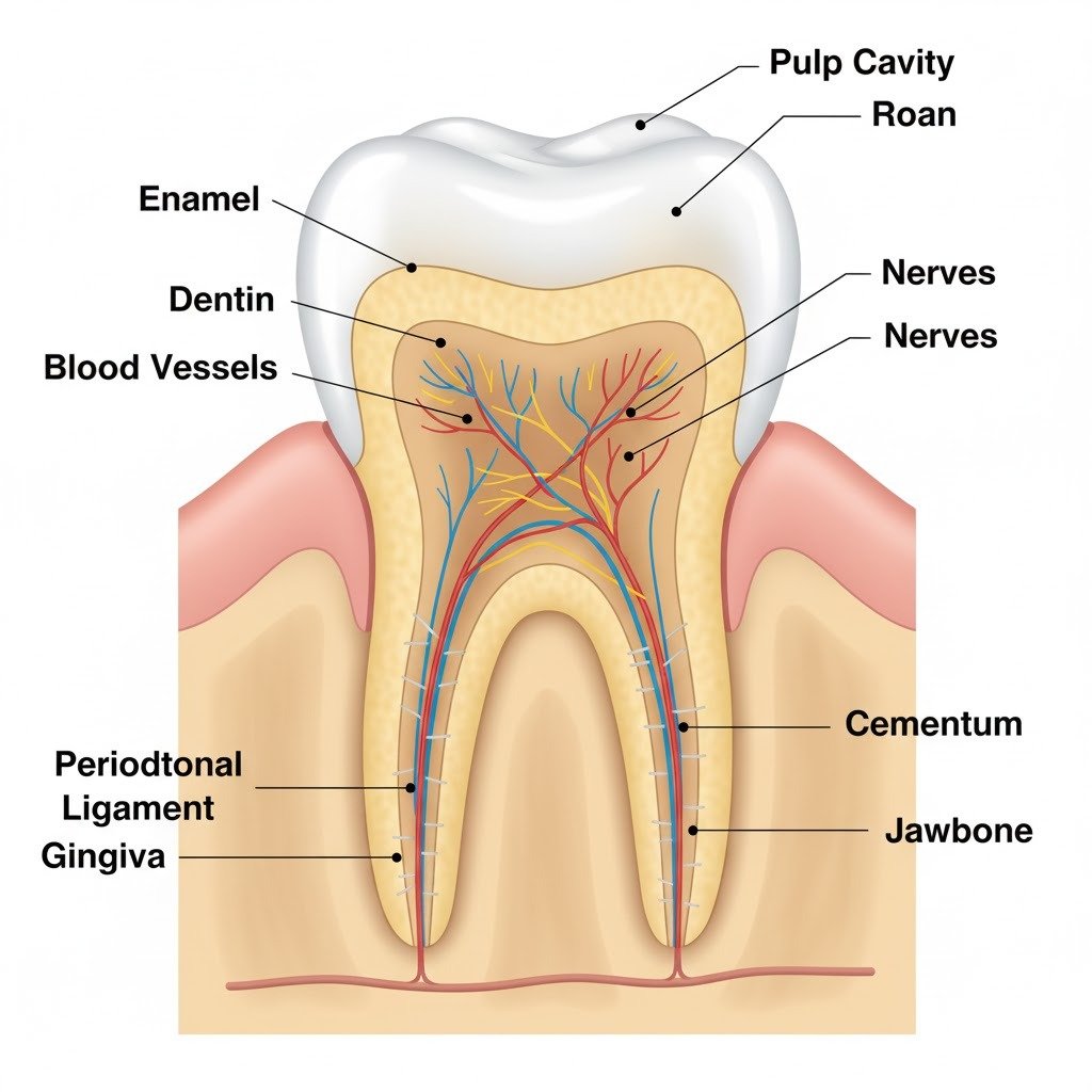
This diagram illustrates the internal structure of a mammalian tooth, showing key components from the crown down to its two roots embedded in the jawbone.
Key Structures Labeled:
- Enamel: The outermost, hardest layer of the tooth, covering the crown.
- Dentin: The layer beneath the enamel and cementum, making up the bulk of the tooth.
- Pulp Cavity: The central chamber of the tooth containing pulp.
- Pulp: Soft tissue within the pulp cavity, containing blood vessels, nerves, and connective tissue.
- Nerves: Sensory fibers running through the pulp.
- Blood Vessels: Supply nutrients to the pulp.
- Cementum: A bone-like tissue covering the root surface.
- Periodontal Ligament: Connects the cementum of the root to the alveolar bone of the jaw.
- Gingiva: The gums, which surround the base of the tooth.
- Jawbone: The bone in which the tooth roots are anchored.
Question 2.
Try to swallow the saliva in your mouth, and feel with your hand your neck. What happens in the neck?
Ans:
When you swallow saliva, you’ll feel a brief upward and downward movement in the front of your neck.
This movement is caused by the rise and fall of your larynx (voice box) and the structure surrounding it, the Adam’s apple (if pronounced).
Mechanism of Swallowing
This movement occurs because swallowing is a complex action designed to ensure food (or saliva) goes down the esophagus and not the windpipe (trachea):
- Laryngeal Elevation: As the swallowing reflex begins, the muscles in the throat pull the larynx (voice box) upward and forward.
- Epiglottis Closure: This upward movement causes the epiglottis (a small flap of cartilage) to fold down and cover the opening of the windpipe (trachea). This seals off the air passage.
- Passage to Esophagus: The closing of the airway directs the saliva (or food/liquid) safely into the esophagus, which then carries it down to the stomach via peristalsis.
- Return to Rest: Once the swallow is complete, the larynx drops back down to its resting position, reopening the airway for breathing.
3. Complete the following table by filling in the blanks 1 to 8.
| Organ | Enzyme | Food acted upon | Find product |
| 1 | Pepsin | 2 | 3 |
| Mouth | 4 | 5 | Disaccharide |
| 6 | 7 | Maltose | 8 |
Ans:
| Organ | Enzyme | Food Acted Upon | Final Product |
| Stomach | Pepsin | 1. Protein | 2. Peptones/Polypeptides |
| Small Intestine | 3. Trypsin/Erepsin | 4. Protein/Peptides | 5. Amino Acids |
| Mouth | 6. Salivary Amylase (Ptyalin) | 7. Starch | 8. Maltose (Disaccharide) |
Explanation of Blanks:
- 1 & 2 (Stomach): Pepsin is the primary enzyme in the stomach. It acts on Protein, breaking it down into smaller polypeptide chains called Peptones and Proteoses.
- 3, 4, & 5 (Small Intestine): Digestion is completed here. Trypsin (from the pancreas) and Erepsin (a group of peptidases from the intestinal wall) act on Protein and peptides, breaking them down into the final absorbable product: Amino Acids.
- 6, 7, & 8 (Mouth): The mouth contains Salivary Amylase. It begins the chemical digestion of Starch, breaking it down into a Disaccharide known as Maltose.
Question 4.
1. Study the diagram given below and then answer the question that follows:

Name the parts labelled 1, 2, 3, 4, 5 and 6.
2. Study the diagram given below and then answer the question that follows:

Identify the tooth and give a reason to support your answer.
3. Study the diagram given below and then answer the question that follows:

Describe the structure of the part labelled ‘3’.
4. Study the diagram given below and then answer the question that follows:
Give the total number of the type of tooth mentioned in ‘1’ above, in the mouth of an adult and state its function.
Ans:
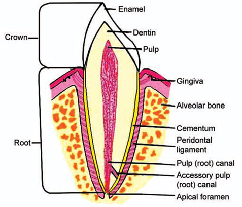
The diagram shows a cross-section of a single-rooted tooth.
Tooth Structure Labeling
Here are the names of the parts labeled in the diagram:
- Enamel
- Dentin
- Pulp Cavity (containing the Pulp, Nerves, and Blood Vessels)
- Gingiva (Gum)
- Crown
- Cementum
2. Identify and Justify the Tooth Type
The tooth is most likely an Incisor or a Canine.
Reason: The diagram shows a tooth with only one root and a relatively simple, chisel-shaped or conical crown (part 5) when compared to the multi-rooted molars and premolars. Incisors (for cutting) and Canines (for tearing) are typically single-rooted.
3. Structure of Part 3 (Pulp Cavity)
Part 3 is the Pulp Cavity (or Pulp Chamber), which houses the Dental Pulp.
The structure of the pulp is a soft, living connective tissue that fills the central cavity of the tooth. It is composed of:
- Blood Vessels: These provide nutrients and oxygen to the surrounding dentin and pulp tissue.
- Nerves: These transmit sensory information, especially pain, to the brain.
- Lymphatics: Vessels that help drain fluid and regulate pressure within the cavity.
- Odontoblasts: Cells that line the outer wall of the pulp cavity and are responsible for forming dentin throughout the tooth’s life.
4. Total Number and Function of Tooth Type
Assuming the tooth shown (single-rooted, simple crown) is an Incisor (part of the group mentioned in the previous analysis):
- Total Number in an Adult Mouth: There are 8 Incisor teeth in the mouth of a human adult (4 in the upper jaw and 4 in the lower jaw).
- Function: The function of the Incisor teeth is cutting and biting food into smaller, manageable pieces.
Question 5.
1.Study the following dental formula and then answer the question that follow:
i 3 / 4 c 0 / 0 pm 0 / 1 m 1 / 1
State the total number of teeth present in the dentition.
2. Study the following dental formula and then answer the question that follow:
i 3 / 4 c 0 / 0 pm 0 / 1 m 1 / 1
Is the dentition that of a carnivore or herbivore? Give a reason to support your answer.
3. Study the following dental formula and then answer the question that follow:
i 3 / 4 c 0 / 0 pm 0 / 1 m 1 / 1
Name an animal possessing such a dentition.
4. Study the Following Dental Formula and Then Answer the Question that Follow:
i 3 / 4 c 0 / 0 pm 0 /1 m 1 / 1
Give the dental formula of an adult human being.
Ans:
1. State the total number of teeth present in the dentition.
To find the total number, we add the numbers on one side of the formula and multiply by 2.
The formula is: i 3/4 c 0/0 pm 0/1 m 1/1
- Incisors (i): 3 (upper) + 4 (lower) = 7
- Canines (c): 0 + 0 = 0
- Premolars (pm): 0 + 1 = 1
- Molars (m): 1 + 1 = 2
- Total for one side: 7 + 0 + 1 + 2 = 10 teeth.
- Total dentition: 10 × 2 = 20 teeth.
2. Is the dentition that of a carnivore or herbivore? Give a reason to support your answer.
This is the dentition of a herbivore.
Reason: The key indicator is the presence of a diastema (a gap) where the canines would be, as shown by the canine formula c 0/0. This gap allows for the manipulation of plant material and helps in the process of chewing cud. Additionally, the incisors are well-developed for cutting and nibbling on vegetation.
3. Name an animal possessing such a dentition.
An animal possessing such a dentition is a Sheep or a Goat. (Other examples include cows and deer).
4. Give the dental formula of an adult human being.
The dental formula of an adult human being is:
i 2/2 c 1/1 pm 2/2 m 3/3
