

The human skeletal system is a dynamic framework composed of bones and cartilage, serving several critical functions beyond just providing shape. It acts as a sturdy scaffold that supports the body’s weight and maintains its posture. Furthermore, it offers crucial protection for delicate internal organs; for instance, the skull encases the brain, and the rib cage shields the heart and lungs. The skeleton is also the body’s primary mineral reservoir, storing calcium and phosphorus, and it is the site for the production of blood cells within the bone marrow. Structurally, the skeleton is divided into the axial skeleton (skull, vertebral column, and rib cage) and the appendicular skeleton, which comprises the limbs and their girdles that connect them to the axial core.
Movement at the joints is a coordinated effort between the skeleton and muscles. Joints are the points where two or more bones meet, and they are classified based on their mobility. Immovable joints, like those in the skull, lock bones together for protection. Slightly movable joints, such as those between vertebrae, allow for limited flexibility. However, the most significant movements are facilitated by freely movable or synovial joints, like the ball-and-socket joint in the shoulder and the hinge joint in the knee. These complex joints are enclosed by a capsule filled with synovial fluid, which acts as a lubricant to reduce friction. The bones are held in place at these joints by tough, fibrous bands of tissue called ligaments, which prevent dislocation and provide stability.
Locomotion, the ability to move from one place to another, is achieved through the integrated work of the skeletal and muscular systems, forming a lever system. This specific interaction is brought about by muscular tissue’s unique property of contractibility. Skeletal muscles, which are attached to bones by tough, inelastic tendons, typically work in antagonistic pairs across a joint. When one muscle, the flexor, contracts to bend a joint, its partner, the extensor, must relax and elongate. This opposing action is clearly seen in the biceps and triceps of the upper arm, which work together to pull the bones and create the coordinated, purposeful movements that allow for walking, running, and other forms of locomotion.
Multiple choice type
Question 1.
Your external ear (pinna) is supported by
- Bone
- Cartilage
- Tendon
- Capsule
Question 2.
The type of joint found at shoulder is also found at
- Elbow
- Knee
- Ankle
- Hip
Question 3.
Which one of the following categories of vertebrae are correctly matched?
- Cervical-7
- Thoracic-10
- Lumbar-4
- Sacral-4
Question 4. The human skeleton altogether contains 213 bones. Which of these are the 6 bones?
- Neck vertebrae
- Ear ossicles
- Carpals
- Metacarpals
Very short answer type
Question 1.
Name the parts of the skeleton where the following are located:
transverse process, glenoid cavity, shoulder-blade, acetabulum
Ans:
| Structure | Location in the Skeleton |
| Transverse Process | Vertebrae (part of the spine/vertebral column) |
| Glenoid Cavity | Scapula (Shoulder Blade) |
| Shoulder-blade | Pectoral Girdle (upper back) |
| Acetabulum | Pelvic Girdle (Hip Bone/Coxal Bone) |
Question 2.
Name any two parts of your body where the supporting skeleton is made of cartilage instead of bone.
Ans:
The supporting skeleton is made of cartilage instead of bone in several parts of your body. Two prominent examples are:
- Outer Ear (Pinna): The entire visible structure of the outer ear is supported by flexible cartilage, allowing it to maintain its shape while remaining pliable.
- Nose Tip: The lower part and tip of your nose are supported by cartilage, giving the nose its shape and flexibility, while the bridge of the nose is bone.
Additional Examples
- Trachea (Windpipe) and Bronchi: The air tubes are kept open by C-shaped rings of cartilage.
- Larynx (Voice Box): Its structure is made almost entirely of cartilage.
- Joints: Cartilage covers the ends of bones at joints (articular cartilage) to reduce friction and cushion movement.
Short answer type
Question 1.
What is the difference between a true rib and a floating rib?
Ans:
| Feature | True Ribs (Vertebrosternal) | Floating Ribs (Vertebral) |
| Number | Rib pairs 1 through 7 (7 pairs, 14 total). | Rib pairs 11 and 12 (2 pairs, 4 total). |
| Anterior Connection | Each rib connects directly to the sternum via its own costal cartilage. | They do not connect to the sternum or to any other cartilage anteriorly. |
| Distal End | Ends are firmly attached and stabilized at the front of the rib cage. | Their cartilaginous ends terminate freely in the muscle of the lateral abdominal wall. |
| Function | Provide the most structural integrity and protection for the heart and lungs. | Provide less protection to the thoracic organs but offer protection to the kidneys. |
Question 2.
Do the muscles pull the structures, or push them? Explain briefly.
Ans:
Muscles can only pull structures; they cannot push them.
Mechanism of Muscle Action
Muscles exert force by contracting (shortening). When a muscle contracts, it pulls the two structures (usually bones) to which it is attached closer together.
- Contraction (Pulling): This active shortening is the only way a skeletal muscle can generate movement. For example, your biceps muscle contracts to pull your forearm toward your shoulder (flexion).
- Relaxation (Inactivity): When a muscle relaxes, it lengthens and becomes passive. It does not actively push the attached structure away.
- Antagonistic Pairs: To move a structure in the opposite direction (e.g., extending your arm), a separate muscle or group of muscles called the antagonist must contract and pull. The movement is always the result of a pulling action by one set of muscles, while the opposing set relaxes.
Question 3.
Just as the humerus corresponds to femur, what bones correspond to tarsals, metacarpals, ulna and radius respectively?
Ans:
| Upper Limb Bone(s) | Lower Limb Correspondence |
| Humerus (Upper Arm) | Femur (Upper Leg/Thigh) |
| Tarsals (Ankle) | Carpals (Wrist) |
| Metacarpals (Palm/Hand) | Metatarsals (Foot) |
| Ulna and Radius (Forearm) | Tibia and Fibula (Lower Leg) |
Question 4.
What are antagonistic muscles? Give one example.
Ans:
Antagonistic muscles are pairs of muscles that work in opposition to one another to produce movement at a joint. When one muscle of the pair contracts (shortens) to pull a bone in one direction, the other muscle of the pair relaxes and lengthens. To move the bone back, the roles reverse.
The muscle that is contracting and causing the movement is called the agonist (or prime mover), and the muscle relaxing is the antagonist.
Example: Biceps and Triceps in the Arm
The classic example of an antagonistic pair is the Biceps and Triceps muscles in the upper arm, which control the movement of the forearm at the elbow joint:
- To Bend the Arm (Flexion): The Biceps muscle contracts (acts as the agonist) and pulls the forearm up, while the Triceps muscle relaxes (acts as the antagonist).
- To Straighten the Arm (Extension): The Triceps muscle contracts (acts as the agonist) and pulls the forearm down, while the Biceps muscle relaxes (acts as the antagonist).
Question 5.
Some people in old age complain of stiff joints. What do you think could be a possible reason for it?
Ans:
Stiff joints in old age are most commonly caused by Osteoarthritis and the general wear and tear of joint structures over decades of use.
Possible Reasons for Stiff Joints
1. Cartilage Degradation (Osteoarthritis)
This is the single most frequent cause. Over time, the articular cartilage—the smooth, protective tissue covering the ends of bones in a joint—wears down.
- Friction: As cartilage thins, bones start rubbing directly against each other, causing pain, inflammation, and stiffness.
- Reduced Cushioning: The loss of cartilage reduces the joint’s ability to absorb shock, leading to restricted, painful movement.
2. Decreased Synovial Fluid
Joints are lubricated by synovial fluid, which reduces friction and provides nourishment.
- Fluid Reduction: With age, the body may produce less synovial fluid, and the fluid itself may become less viscous (thinner).
- Stiffness: Reduced lubrication increases friction, leading to a stiff and creaky feeling, especially after periods of inactivity (e.g., first thing in the morning).
3. Ligament and Tendon Changes
The connective tissues surrounding the joint also change with age.
- Loss of Elasticity: Ligaments and tendons become less elastic and more rigid due to changes in their collagen structure.
- Reduced Flexibility: This makes the joint capsule tighter, restricting the full range of motion and contributing to the feeling of stiffness.
Long answer type
Question 1.
What are the uses of the skeleton in our body?
Ans:
The skeleton is far more than a simple structural frame; it is a dynamic system integral to numerous bodily processes.
Essential Functions of the Human Skeleton
1. Structural Scaffolding
The bony framework provides support and shape to the body. By countering the pull of gravity, the skeleton allows us to maintain an upright posture and serves as the anchorage point for all soft tissues and organs.
2. Safeguarding Internal Organs
Specific bony enclosures are dedicated to protecting vital, delicate organs:
- The skull acts as a hard casing for the brain.
- The rib cage and breastbone shield the heart and lungs.
- The column of vertebrae encases and protects the spinal cord.
3. Facilitating Locomotion
Bones function as rigid levers. Skeletal muscles attach to these levers via tendons, and when the muscles contract, they pull on the bones. Joints provide the movable connections that allow these pulling forces to translate into a vast range of body movements and locomotion.
4. Mineral Reserves
Bones act as the body’s major reservoir for crucial inorganic salts, most notably calcium and phosphorus. These minerals are stored and released into the bloodstream as needed, helping to regulate their levels, which are critical for nerve impulse transmission, muscle contraction, and blood clotting.
5. Production of Blood Components
The spongy tissue found inside certain large bones, known as red bone marrow, is the site of hematopoiesis. This essential process is where the body continuously manufactures all its blood elements: red blood cells, white blood cells, and platelets.
Question 2.
What are the different types of joints? Give one example of each type.
Ans:
Joints, or articulations, are classified based on the degree of movement they allow. The three main functional types of joints are fibrous, cartilaginous, and synovial.
Types of Joints and Examples
1. Immovable (Fibrous) Joints (Synarthroses)
These joints are held together by dense fibrous connective tissue, allowing no movement at all. They are essential for protecting vital organs.
- Structure: Bones are interlocked or joined by a short ligament.
- Example: Sutures found between the bones of the skull (e.g., between the parietal and frontal bones).
2. Slightly Movable (Cartilaginous) Joints (Amphiarthroses)
These joints are united by cartilage (either hyaline or fibrocartilage), which allows for limited movement in response to pressure or bending.
- Structure: Bones are connected by a plate of cartilage.
- Example: The joints between the vertebrae (connected by intervertebral discs of fibrocartilage) or the pubic symphysis (joining the two hip bones).
3. Freely Movable (Synovial) Joints (Diarthroses)
These are the most common and structurally complex joints. They are characterized by a joint cavity filled with synovial fluid, which acts as a lubricant, allowing for a wide range of motion.
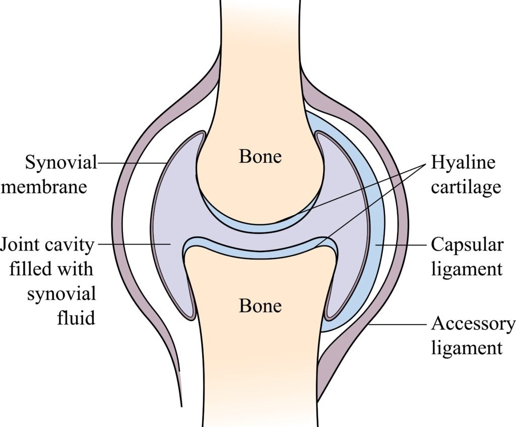
- Structure: Articular cartilage covers the bone ends, and the joint is enclosed by a fibrous capsule lined with a synovial membrane.
- Example: The Knee Joint (a hinge joint) or the Shoulder Joint (a ball-and-socket joint).
Question 3.
What is the difference between ligament and tendon? What are their functions?
Ans:
Ligament vs. Tendon
| Feature | Ligament | Tendon |
| Connection | Connects bone to bone. | Connects muscle to bone. |
| Tissue Composition | More elastic fibers, allowing for a degree of stretch. | Less elastic, very strong collagen fibers. |
| Appearance | Often shorter and band-like. | Cord-like or sheet-like. |
Functions
Ligament Functions
Ligaments primarily serve a stabilizing and limiting function at joints.
- Joint Stability: They hold bones together in a joint, preventing them from separating or dislocating.
- Limit Movement: They restrict the range of motion of the joint, ensuring it moves only in its intended direction and preventing hyperextension or excessive rotation.
Tendon Functions
Tendons primarily serve a kinetic (movement) function.
- Transmit Force: They act as strong cables, transmitting the mechanical force generated by muscle contraction to the bone.
- Facilitate Movement: By pulling on the bone, tendons facilitate the movement of the skeleton across the joint, allowing for activities like walking, lifting, and running.
Question 4.
What are bones made of? Are the bones living or non living? Give reasons.
Ans:
Bones are complex structures primarily made of a combination of organic and inorganic materials. They are definitely living tissues.
Composition of Bones
Bones have two main components that provide their unique properties:
- Inorganic Component (Hardness): About two-thirds of the bone’s weight consists of mineral salts, mainly calcium phosphate and calcium carbonate (hydroxyapatite). This mineral matrix provides the bone’s hardness and rigidity.
- Organic Component (Flexibility): About one-third of the bone is made of organic material, primarily collagen fibers (a protein) and various cells. This component provides the bone’s flexibility and tensile strength, preventing it from shattering easily.
Are Bones Living or Non-Living?
Bones are living tissues (living organs) for several critical reasons:
- Contain Living Cells: Bones are composed of several types of specialized, living cells:
- Osteoblasts: Build new bone tissue.
- Osteoclasts: Break down old bone tissue.
- Osteocytes: Mature bone cells that maintain the bone matrix.
- Active Metabolism: Bone tissue requires a constant supply of blood, oxygen, and nutrients (like Calcium and Vitamin D) to perform metabolic functions.
- Growth and Repair: Bones can grow, change shape throughout life in response to stress (remodeling), and repair themselves after a fracture, all of which are characteristics of living tissue.
- Innervation: Bones contain nerves and are therefore sensitive to pain.
Question 5.
Below is a diagram of the human skeleton. Name the bones numbered 1-11.
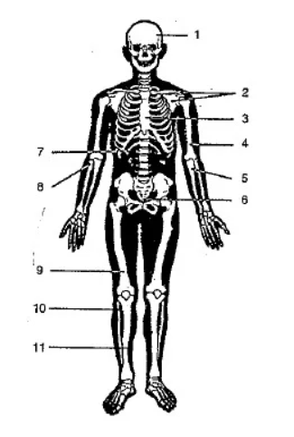
Ans:
| Number | Name of Bone |
| 1 | Skull (or Cranium) |
| 2 | Clavicle (Collarbone) |
| 3 | Sternum (Breastbone) |
| 4 | Humerus (Upper arm bone) |
| 5 | Radius (Lateral forearm bone) |
| 6 | Pelvic Girdle (Hip bone) |
| 7 | Ribs (Rib cage) |
| 8 | Ulna (Medial forearm bone) |
| 9 | Femur (Thigh bone) |
| 10 | Patella (Kneecap) |
| 11 | Tibia (Shin bone) |
