

The respiratory system is fundamentally designed for the essential process of gas exchange. Its primary function is to supply our body’s cells with oxygen, which is crucial for breaking down food to release energy, a process called cellular respiration. Simultaneously, it must remove the waste product carbon dioxide from the body. This entire process, known as breathing or ventilation, involves two main phases: inhalation, where air rich in oxygen is drawn into the lungs, and exhalation, where air laden with carbon dioxide is forced out. The journey of air begins through the nostrils, where it is filtered by nasal hairs, warmed, and moistened before traveling through the pharynx, larynx (or voice box), and into the trachea.
The trachea, or windpipe, is a sturdy tube kept open by rings of cartilage. It branches into two smaller tubes called bronchi, one leading to each lung. Inside the lungs, the bronchi further divide into a vast network of finer and finer tubes known as bronchioles, resembling the branches of an upside-down tree. This intricate network finally ends in millions of tiny, air-filled sacs called alveoli. The alveoli are the true sites of gas exchange; their thin, moist walls are surrounded by a dense web of capillaries. This structure creates an immense surface area, making the diffusion of gases highly efficient. Oxygen from the inhaled air dissolves in the moisture and diffuses into the blood, while carbon dioxide from the blood diffuses out into the alveolar air to be exhaled.
Breathing is an involuntary action controlled by the respiratory center in the brain, specifically the medulla oblongata. The physical act of breathing is driven by the movement of the diaphragm—a dome-shaped muscle at the base of the chest—and the intercostal muscles between the ribs. During inhalation, the diaphragm flattens and contracts while the rib cage moves upward and outward, increasing the chest volume and pulling air into the lungs. Exhalation is typically a passive process where these muscles relax, the chest cavity volume decreases, and air is pushed out. Several factors can affect our breathing rate, including physical activity, emotional state, and the levels of carbon dioxide in the blood, which the brain constantly monitors.
Multiple choice type
Question 1.
During inspiration, the diaphragm
- relaxes
- contracts
- expands
- gets folded
Question 2.
The ultimate end parts of the respiratory system in humans are known as
- alveoli
- bronchioles
- tracheoles
- Bronchi
Question 3.
During respiration there is
- gain in dry weight
- loss in dry weight
- no change in dry weight
- increase in the overall weight
Very short answer type
Question 1.
1. Choose the odd one out in the following groups of four items each: Trachea, Bronchus, Alveolus, Diaphragm
2. Choose the odd one out in the following groups of four items each: Ethyl alcohol, Carbon dioxide, Starch, Oxygen
3. Choose the odd one out in the following groups of four items each: Diffusion, Respiratory gases, Alveoli, Capillary network
4. Choose the odd one out in the following groups of four items each: Trachea, Ciliated epithelium, Mucous, Diffusion
5. Choose the odd one out in each of the following groups of four items each:
Oxyhaemoglobin, Carbaminohaemoglobin, Hypoxia, Carboxyhaemoglobin
6. Choose the odd one out in the following groups of four items each: Hairy, Moist, Nostril, Vocal cord
Ans:
Here are the odd ones out from each group, along with a brief explanation:
1. Trachea, Bronchus, Alveolus, Diaphragm
The odd one out is Diaphragm.
- Reason: The Trachea, Bronchus, and Alveolus are all hollow air-conducting or gas-exchange structures within the respiratory tract. The Diaphragm is a sheet of muscle that powers the breathing movements.
2. Ethyl alcohol, Carbon dioxide, Starch, Oxygen
The odd one out is Starch.
- Reason: Ethyl alcohol, Carbon dioxide, and Oxygen are all directly involved as products or reactants in the process of respiration (both anaerobic and aerobic). Starch is a complex carbohydrate that serves as a fuel source but is not a direct reactant or product in the final respiratory pathways.
3. Diffusion, Respiratory gases, Alveoli, Capillary network
The odd one out is Diffusion.
- Reason: Alveoli, Respiratory gases (O2,CO2), and the Capillary network are all physical structures or substances involved in the gas exchange process. Diffusion is the process (mechanism) itself by which the gases move.
4. Trachea, Ciliated epithelium, Mucous, Diffusion
The odd one out is Diffusion.
- Reason: Trachea, Ciliated epithelium, and Mucous are all related to the filtering and conducting function of the major airways (trachea and bronchi). Diffusion is the process of gas exchange that occurs only in the alveoli, not typically in the conducting airways.
5. Oxyhaemoglobin, Carbaminohaemoglobin, Hypoxia, Carboxyhaemoglobin
The odd one out is Hypoxia.
- Reason: Oxyhaemoglobin, Carbaminohaemoglobin, and Carboxyhaemoglobin are all compounds formed when haemoglobin binds chemically with a gas (O2,CO2,CO respectively). Hypoxia is a physiological state defined as a deficiency in the amount of oxygen reaching the tissues.
6. Hairy, Moist, Nostril, Vocal cord
The odd one out is Vocal cord.
- Reason: Hairy, Moist, and Nostril are all characteristics or features associated with the initial filtration and conditioning of air as it enters the nasal passage. The Vocal cord is a structure in the larynx involved in sound production and is not primarily involved in air conditioning or filtration.
Question 2.
1.Name the body structure concerned with the given functional activity: Prevents food from entering the trachea during swallowing.
2. Name the body structure concerned with the given functional activity: Transports oxygen to the body cells.
3. Name the body structure concerned with the given functional activity: Helps to increase the volume of the chest cavity lengthwise.
4. Name the body structure concerned with the given functional activity: Combined with the oxygen in the lungs.
5. Name the body structure concerned with the given functional activity: Protects the lungs from mechanical injury
6.Name the body structure concerned with the given functional activity: Provides actual diffusion of respiratory gases in the lungs.
Ans:
Here are the structural components of the human body responsible for the specified physiological activities, presented with original phrasing and emphasis on the core function:
1. Airway Protection
The structure responsible for ensuring that ingested material does not enter the windpipe during the act of deglutition (swallowing) is the Epiglottis. This cartilaginous flap seals the entrance to the larynx and trachea when food passes, directing it safely toward the esophagus.
2. Oxygen Delivery System
The component tasked with moving oxygen from the pulmonary system to the body’s tissues is the Blood, specifically the hemoglobin molecules contained within the red blood cells. Hemoglobin serves as the primary carrier molecule for molecular oxygen.
3. Vertical Chest Expansion
The principal muscle that facilitates the increase in the vertical dimension of the thoracic cavity (chest) during the breathing cycle is the Diaphragm. Its contraction, drawing it downward, is essential for inspiration (inhalation).
4. Oxygen Binding Agent
The specialized protein that reversibly associates with oxygen within the capillaries of the lungs is Hemoglobin. This iron-containing protein is responsible for the crucial exchange of gases at the respiratory surface.
5. Thoracic Protection
The skeletal framework that shields the pulmonary organs from external force and injury is the Rib Cage (composed of the Ribs and Sternum).
6. Gas Exchange Site
The tiny, thin-walled air sacs where the actual exchange of respiratory gases (O2 and CO2) occurs in the lungs is the Alveoli. They provide the immense surface area necessary for efficient diffusion.
Question 3.
What is the normal percentage composition of gases in inspired and expired air respectively?
Ans:
| Gas | Inspired Air (Inhaled) | Expired Air (Exhaled) | Reason for Change |
| Nitrogen (N2) | ~78.09% | ~78.09% | Nitrogen is an inert gas and is not used by the body. |
| Oxygen (O2) | ~21.00% | ~16.00% | Oxygen is consumed by the body for cellular respiration. |
| Carbon Dioxide (CO2) | ~0.04% | ~4.00% | Carbon Dioxide is produced as a waste product of cellular respiration and released. |
| **Water Vapor (H2O) ** | Varies (Low/Variable) | ~6.00% (Saturated) | Air is humidified by the warm, moist respiratory tract before being exhaled. |
Question 4.
Which chemical compound inside a cell can be termed “Currency of Energy”?
Ans:
The chemical compound inside a cell that can be termed the “Currency of Energy” is Adenosine Triphosphate (ATP) .
Why ATP is the “Currency”
ATP is called the energy currency because:
- It is the immediate and universal source of usable energy for almost all cellular activities, such as muscle contraction, active transport, and biosynthesis.
- When a cell needs energy, it breaks this terminal bond to release energy, converting ATP to Adenosine Diphosphate (ADP).
- Like a currency, ATP is constantly “spent” (hydrolyzed to ADP) in reactions that require energy and constantly “earned” (synthesized back from ADP) through processes like cellular respiration.
Question 5.
Match the items in Column I with the ones most appropriate in Column II. Rewrite the matching pairs.
| Column I | Column II |
| (a) Alveoli | (i) where aerobic respiration takes place |
| (b) Bronchioles | (ii) lined with hair |
| (c) Nasal Chamber | (iii) diffusion of gases |
| (d) Bronchi | (iv) small air tubes |
| (v) an inverted Y shaped tube | |
| (vi) a common passage for food and air |
Ans:
| Column I | Column II | Matching Pair |
| (a) Alveoli | (iii) diffusion of gases | Alveoli – diffusion of gases |
| (b) Bronchioles | (iv) small air tubes | Bronchioles – small air tubes |
| (c) Nasal Chamber | (ii) lined with hair | Nasal Chamber – lined with hair |
| (d) Bronchi | (v) an inverted Y shaped tube | Bronchi – an inverted Y shaped tube |
Short answer type
Question 1.
1.Given below is an example of certain structure and its special functional activity:
“Kidney and excretion”. Fill in the blanks on a similar pattern. Alveoli and _____________. 2. Given below is an example of certain structure and its special functional activity:
“Kidney and excretion”. Fill in the blanks on a similar pattern. Mitochondria and _____________.
3. Given below is an example of a certain structure and its special functional activity: “Kidney and excretion”. Fill in the blanks on a similar pattern. Epiglottis and _____________.
4. Given below is an example of a certain structure and its special functional activity:
“Kidney and excretion”. Fill in the blanks on a similar pattern. Pleura and _____________. 5. Given below is an example of certain structure and its special functional activity:
“Kidney and excretion”. Fill in the blanks on a similar pattern. Diaphragm and _____________.
6. Given below is an example of a certain structure and its special functional activity: “Kidney and excretion”. Fill in the blanks on a similar pattern. ‘C’ shaped cartilage rings and ____________.
Ans:
Here are the completions following the pattern “Structure and special functional activity”:
- Alveoli and Gas exchange
- Mitochondria and Cellular respiration (or ATP production)
- Epiglottis and Closing of windpipe (or Preventing food entry into trachea)
- Pleura and Protection (or Lubrication)
- Diaphragm and Breathing (or Inspiration/Respiration)
- ‘C’ shaped cartilage rings and Preventing collapse of trachea (or Support)
Question 2.
1. State one function of the following: Ciliated epithelium lining the respiratory tract
2. State one function of the following: Mitochondria
3. State one function of the following:Diaphragm
4. State one function of the following: Intercostal muscles
5. State one function of the following: Pleural fluid
Ans:
Here is one main function for each of the listed biological structures:
- Ciliated epithelium lining the respiratory tract: To sweep mucus and trapped dust/foreign particles up and out of the respiratory passages (the mucociliary escalator), preventing them from reaching the lungs.
- Mitochondria: To generate the majority of the cell’s supply of usable energy in the form of Adenosine Triphosphate (ATP) through cellular respiration.
- Diaphragm: To facilitate inhalation (breathing in) by contracting and flattening, which increases the vertical volume of the chest cavity.
- Intercostal muscles: To move the ribs upward and outward during inhalation, and inward during forced exhalation, thereby changing the volume of the chest cavity.
- Pleural fluid: To lubricate the opposing surfaces of the lungs and the chest wall (the two layers of the pleura), allowing the lungs to slide smoothly against the thoracic wall during breathing without friction.
Question 3.
Match the items in Column A with those in Column B.
| Column A | Column B |
| Cartilaginous | Epiglottis |
| Large surface area | Diaphragm |
| Breathing movements | Bronchi |
| Voice | Alveoli |
| Complemental air | Larynx |
| Swallowing | Extra inhalation |
Ans:
| Column A | Column B | Matching Pair |
| Cartilaginous | Larynx | Cartilaginous – Larynx |
| Large surface area | Alveoli | Large surface area – Alveoli |
| Breathing movements | Diaphragm | Breathing movements – Diaphragm |
| Voice | Larynx | Voice – Larynx |
| Complemental air | Extra inhalation | Complemental air – Extra inhalation |
| Swallowing | Epiglottis | Swallowing – Epiglottis |
Explanations:
- Larynx (Voice Box) is primarily composed of cartilage and houses the vocal cords responsible for voice production.
- Alveoli have a structure providing a large surface area for efficient gas exchange.
- The Diaphragm is the primary muscle responsible for driving breathing movements.
- Complemental air is the volume of air that can be inhaled additionally after a normal inspiration (i.e., extra inhalation).
- The Epiglottis is the flap that closes over the windpipe during swallowing to prevent food from entering the trachea.
Question 4.
Under what conditions would the breathing rate increase?
Ans:
The breathing rate (respiratory rate) increases when the body needs to take in more oxygen or eliminate more carbon dioxide. This is primarily controlled by chemoreceptors that monitor the levels of gases in the blood.
Here are the main conditions under which breathing rate increases:
- Increased Physical Activity (Exercise) :
- Muscles perform more cellular respiration, rapidly using upO2 and producing large amounts of CO2.
- The resulting increase in blood CO2 concentration (which lowers the blood pH) is the primary stimulus that signals the respiratory center in the brain to increase the depth and frequency of breathing.
- High Altitude :
- At high altitudes, the partial pressure of oxygen in the air is lower.
- This reduced O2 pressure leads to a lower concentration of O2 in the blood, prompting the respiratory center to increase the breathing rate to compensate and maximize O2 intake.
- Fever or Increased Body Temperature:
- A rise in body temperature increases the overall metabolic rate of cells. This means more O2 is needed and more CO2 is produced, leading to faster breathing.
- Emotional Stress or Excitement:
- Strong emotions like fear, anxiety, or excitement trigger the sympathetic nervous system, leading to involuntary, rapid, and often shallow breathing.
- Acidosis:
- Any medical condition (like untreated diabetes, causing ketoacidosis) that leads to an excessive buildup of acids in the blood will cause the breathing rate to increase dramatically (often called Kussmaul breathing) as the body attempts to expel CO2 to raise the blood pH.
Question 5.
How would you prove that the air you breathe out is warmer?
Ans:
Proving Exhaled Air is Warmer
Method 1: Using a Thermometer
This is the most direct and quantitative method:
- Measure Ambient Air: Use a sensitive laboratory thermometer (or a digital thermometer) to measure the temperature of the room air (the air you breathe in).
- Measure Exhaled Air: Place the bulb or sensor of the same thermometer just in front of your mouth. Exhale slowly and steadily directly over the sensor without touching it.
- Compare: You will observe that the thermometer reading increases, showing that the temperature of the expired air is higher than the initial room (inspired) air.
Method 2: Using the Condensation Principle
This method works well when the ambient air is cool:
- Use a Cool Surface: Find a piece of smooth, cool material, such as a clean, dry mirror, a cold piece of glass, or a polished metal surface.
- Exhale on the Surface: Hold the surface close to your mouth and exhale gently onto it.
- Observation: Moisture (water vapor) from your breath will instantly condense on the cool surface, forming a visible fog or mist.
- Conclusion: The moisture comes from the lungs where the air has been warmed to body temperature (around $37^\circ\text{C}$) and fully saturated with water vapor. When this warm, moist air meets the cooler surface, it loses heat, and the water vapor turns into liquid droplets, proving the breath was significantly warmer than the object’s surface temperature.
Question 6.
How is the respiratory passage kept free of dust particles?
Ans:
The respiratory passage is kept remarkably free of dust particles and foreign debris through a sophisticated set of defensive mechanisms, primarily involving filtration, trapping, and removal.
Mechanisms for Cleaning the Airways
1. Filtration (Nasal Cavity)
The first line of defense is in the nasal passage:
- Hairs (Cilia): The nostrils are lined with coarse hairs that act as a physical filter, trapping large dust particles and foreign matter before the air proceeds deeper into the system.
2. Trapping (Mucous Membrane)
The air is conditioned and cleaned as it moves through the trachea and bronchi:
- Mucous Layer: The entire inner lining of the nasal passage, trachea, and bronchi is covered by a sticky layer of mucous secreted by specialized cells. This mucous effectively traps fine dust, pollen, smoke particles, and bacteria.
3. Removal (Ciliary Action)
Once trapped, the particles are actively removed by the cilia:
- Ciliated Epithelium: The cells lining the trachea and bronchi are covered with tiny, hair-like projections called cilia.
- Mucociliary Escalator: These cilia beat in a synchronized, wave-like motion, creating an upward current that continuously sweeps the mucous layer (and the trapped particles) up toward the pharynx (throat).
- Expulsion: When the mucous reaches the throat, it is either swallowed (where the germs are neutralized by stomach acid) or coughed/sneezed out.
Question 7.
What is wrong in the statement “We breathe in oxygen and breathe out carbon dioxide”.
Ans:
Here is the breakdown of why the statement is inaccurate:
Inaccurate Representation
1. Inspired Air is a Mixture
When we inhale, we breathe in ambient air, which is primarily composed of:
- Nitrogen (N2): Approximately 78%
- Oxygen (O2): Approximately 21%
- Other Gases (including CO2): Less than 1%
The body only uses a portion of the inhaled oxygen; the large amount of nitrogen and the remaining oxygen are simply exhaled.
2. Expired Air is Also a Mixture
When we exhale, the air we breathe out is the result of gas exchange in the lungs and is also a complex mixture:
- Nitrogen (N2): Remains high (around 78%)
- Oxygen (O2): Still present, but reduced (around 16%)
- Carbon Dioxide (CO2): Increased significantly (around 4%)
- Water Vapor (H2O): High (saturated)
Conclusion: We do not breathe only oxygen and do not breathe out only carbon dioxide. Breathing is the process of exchanging air mixtures, where the body’s only goal is to reduce the percentage of oxygen and increase the percentage of carbon dioxide.
Long answer type
Question 1.
1. Differentiate between the following pairs on the basis of the aspect given in the brackets. Aerobic and Anaerobic respiration (End products of the process)
2. Differentiate between the following pairs on the basis of the aspect given in the brackets. Respiration and Photosynthesis (Gas released)
3. Differentiate between the following pairs on the basis of the aspect given in the brackets. Photosynthesis and Respiration (Reactants)
4. Differentiate between the following pairs on the basis of the aspect given in the brackets. Inspired air and Alveolar air (CO2 content)
5. Differentiate between the following pairs on the basis of the aspect given in the brackets. Respiration and Breathing (Organs involved)
6. Differentiate between the following pairs on the basis of the aspect given in the brackets. Tidal volume and Residual volume (Quantity of air)
Ans:
1. Aerobic and Anaerobic Respiration (End Products of the Process)
| Process | End Products |
| Aerobic Respiration | Carbon Dioxide (CO2) and Water (H2O), plus a high amount of energy (ATP). |
| Anaerobic Respiration | Lactic Acid (in muscles) OR Ethanol (C2H5OH) and Carbon Dioxide (CO2) (in yeast/plants), plus a low amount of energy (ATP). |
2. Respiration and Photosynthesis (Gas Released)
| Process | Gas Released |
| Respiration | Carbon Dioxide (CO2) |
| Photosynthesis | Oxygen (O2) |
3. Photosynthesis and Respiration (Reactants)
| Process | Reactants |
| Photosynthesis | Carbon Dioxide (CO2) and Water (H2O) (with light energy). |
| Respiration | Glucose (C6H12O6) and Oxygen (O2). |
4. Inspired air and Alveolar air (CO2 content)
| Air Type | CO2 Content |
| Inspired Air | Very low (≈0.04%), reflecting the ambient atmosphere. |
| Alveolar Air | Much higher (≈5.3% or 100 times more) due to the CO2 continuously diffusing out of the blood into the alveoli. |
5. Respiration and Breathing (Organs Involved)
| Process | Organs Involved |
| Respiration | Mitochondria (within every living cell) and cytoplasm, where gas exchange actually occurs. |
| Breathing | Respiratory organs like the lungs, trachea, bronchi, rib cage, and diaphragm, which facilitate the mechanical movement of air. |
6. Tidal volume and Residual volume (Quantity of air)
| Volume Type | Quantity of Air |
| Tidal Volume | The volume of air inhaled or exhaled in a single normal, quiet breath (≈500 mL). |
| Residual Volume | The volume of air that always remains in the lungs even after the most forceful exhalation (≈1200 mL). |
Question 2.
1. Give Suitable Explanation for the Following : Breathing through the nose is said to be healthier than through the mouth.
2. Give a suitable explanation for the following : Why does gaseous exchange continue in the lungs even during expiration?
3. Give a suitable explanation for the following : Why does a person feel breathlessness at higher altitudes?
4. Give a suitable explanation for the following : Why do you shiver and why do your teeth chatter when it is very cold in winter?
Ans:
1. Breathing Through the Nose vs. Mouth
Breathing through the nose is healthier than through the mouth because the nasal passage is equipped with specialized features to filter, warm, and humidify the inspired air before it reaches the delicate lungs:
- Filtration: The nose hairs and sticky mucus trap dust, pollen, and large germs, preventing them from entering the respiratory tract.
- Warming: Blood capillaries lining the nasal cavity quickly heat the incoming air close to body temperature, protecting the lung tissues.
- Humidification: The moist mucosal lining saturates the air with water vapor, preventing the drying out of the tracheal and bronchial membranes.
Breathing through the mouth bypasses these crucial conditioning and filtering mechanisms, sending cold, dry, and dirty air directly to the lungs.
2. Gaseous Exchange During Expiration
Gaseous exchange (oxygen into the blood, carbon dioxide out of the blood) continues in the lungs even during expiration because the process relies on diffusion, not on the physical movement of air.
- Continuous Concentration Gradient: During the brief pause and the process of exhalation, the concentration gradient for the gases still exists across the alveolar walls. The blood arriving at the lungs still contains less O2 and more CO2 than the air remaining in the alveoli.
- Efficient Diffusion: As long as there is a difference in the partial pressures of the gases, diffusion will continue until equilibrium is reached or until the next breath disrupts the balance. This ensures the blood is optimally saturated with oxygen.
3. Breathlessness at Higher Altitudes
A person feels breathlessness (altitude sickness or hypoxia) at higher altitudes because the partial pressure of oxygen (O2) decreases significantly, even though the percentage of oxygen in the air remains the same (21%).
- Lower Atmospheric Pressure: At high altitudes, the total atmospheric pressure is much lower.
- Reduced O2 Driving Force: Because the total pressure is lower, the partial pressure of O2 is also lower. This means there is a weaker driving force (a smaller pressure gradient) to push oxygen from the alveoli into the blood, making the diffusion process less efficient.
- Hypoxia: The body senses this decreased oxygen loading in the blood, triggering a reflex to breathe faster and deeper to compensate, leading to the feeling of breathlessness.
4. Shivering and Teeth Chattering in Cold
You shiver and your teeth chatter when it is very cold in winter because these are involuntary muscular responses designed to generate heat and raise your falling core body temperature (thermoregulation).
- Shivering: This is the rapid, rhythmic, involuntary contraction of skeletal muscles. Since all muscle contraction requires energy and produces a large amount of heat as a byproduct, shivering is an extremely effective way to quickly raise the body’s internal heat production (thermogenesis).
- Teeth Chattering: This is a specific form of shivering involving the rapid, involuntary contractions of the jaw muscles, which are among the most powerful muscles in the body, thus maximizing heat production in the head and neck area.
Question 3.
1. With regard to the respiratory system and the process of respiration in man, answer the following questions: Name the two muscles that help in breathing.
2. With regard to the respiratory system and the process of respiration in man, answer the following question: Briefly describe how the above mentioned muscles help in the inspiration of air.
3. With regard to the respiratory system and the process of respiration in man, answer the following question : Give the overall chemical equation to represent the process of respiration in humans.
4. With regard to the respiratory system and the process of respiration in man, answer the following question: What is meant by : Residual air and Dead air space
Ans:
1. Muscles of Breathing
The two main muscles that help in breathing are the Diaphragm and the Intercostal Muscles.
2. Inspiration Mechanism
Inspiration (inhalation) is an active process involving the contraction of these two muscle groups:
- Diaphragm: This dome-shaped muscle located beneath the lungs contracts and flattens, moving downwards. This action significantly increases the vertical volume of the chest cavity.
- External Intercostal Muscles: These muscles, located between the ribs, contract and pull the ribs upward and outward. This increases the volume of the chest cavity both sideways and front-to-back.
The overall increase in chest volume lowers the air pressure inside the lungs below the atmospheric pressure outside, creating a pressure gradient. This pressure difference forces air to rush into the lungs.
3. Chemical Equation for Respiration
The overall chemical equation to represent the process of aerobic respiration in humans (and most living organisms) is:
This equation shows that glucose and oxygen are used to produce carbon dioxide, water, and energy.
4. Residual Air and Dead Air Space
Residual Air (or Residual Volume)
Residual air is the volume of air that always remains in the lungs and respiratory passages even after the most forceful exhalation. It cannot be expelled. Its function is to keep the lungs inflated and prevent the air sacs (alveoli) from collapsing.
Dead Air Space
Dead air space is the volume of air that fills the respiratory passages (nose, pharynx, larynx, trachea, bronchi, and bronchioles) but does not reach the alveoli where gas exchange occurs. This air is merely transported back and forth; it is considered “dead” because it plays no role in the diffusion of oxygen and carbon dioxide.
Question 4.
Starting from the nostrils, trace the path in sequence which the inspired air takes until it reaches the air sacs.
Ans:
Starting from the nostrils, the inspired air follows a specific sequence of structures that filter, warm, and humidify it until it reaches the air sacs (alveoli) where gas exchange occurs.
Path of Inspired Air
The air follows this sequence from the outside environment to the deep parts of the lungs:
- Nostrils (External Nares): The entry points where air is first filtered by coarse hairs.
- Nasal Chambers/Cavity: The air passes through this space, where it is warmed by blood capillaries and humidified by mucus.
- Pharynx (Throat): A common passage for both air and food; air passes into the lower part.
- Larynx (Voice Box): Air passes through this cartilaginous structure, which contains the vocal cords.
- Trachea (Windpipe): A tube held open by C-shaped cartilage rings. Air travels down this main airway.
- Bronchi (Singular: Bronchus): The trachea divides into two primary bronchi—one leading to each lung.
- Bronchioles: The bronchi continue to divide and branch out into progressively smaller tubes within the lung tissue.
- Alveolar Ducts: The bronchioles terminate in very fine ducts leading to the air sacs.
- Alveoli (Air Sacs): The ultimate end structures where the air finally reaches and where the exchange of oxygen and carbon dioxide takes place with the surrounding capillary network.

Question 5.
1. What are the functions of the following in breathing? Ribs
2. What are the functions of the following in breathing? Diaphragm
3. What are the functions of the following in breathing? Abdominal muscles
Ans:
Here are the functions of the specified body structures in the process of breathing:
1. Ribs
The ribs, along with the sternum (breastbone), form the rib cage. Their primary functions in breathing are:
- Protect the Lungs: They provide a rigid, protective casing for the lungs and heart.
- Increase Chest Volume (Inhalation): The external intercostal muscles attached to the ribs contract, pulling the ribs upward and outward. This increases the chest cavity’s volume, lowering internal pressure and drawing air into the lungs.
2. Diaphragm
The diaphragm is the large, dome-shaped sheet of muscle located beneath the lungs.
- Primary Muscle of Respiration: It is the most important muscle for normal, quiet breathing.
- Increase Chest Volume (Inhalation): When it contracts, the dome flattens and moves downward. This significantly increases the vertical volume of the chest cavity, initiating inhalation.
- Decrease Chest Volume (Exhalation): When it relaxes, it returns to its dome shape and moves upward, pushing air out passively during quiet exhalation.
3. Abdominal Muscles
The abdominal muscles (e.g., rectus abdominis and obliques) are not active during quiet breathing but are essential for forced or deep breathing.
- Forceful Exhalation: They contract powerfully, pushing the abdominal organs and the diaphragm sharply upward. This greatly increases the pressure inside the chest cavity, forcing a large volume of air rapidly out of the lungs (e.g., when coughing, shouting, or during heavy exercise).
Structured/Application/Skill type
Question 1.
1. Given alongside is a diagrammatic sketch of a kind of part in human lungs. Name the parts numbered 1-4
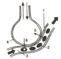
2. Given alongside is a diagrammatic sketch of a kind of part in human lungs.
What do the arrows 5 and 6 indicate?

Ans:
Based on the diagram, which illustrates gas exchange in the lungs:
1. Naming the Numbered Parts
The numbers correspond to the structures involved in oxygen and carbon dioxide exchange:
- Bronchiole (or Alveolar Duct/Terminal Bronchiole) – The small air tube leading into the air sac.
- Capillary (Blood Vessel) – The tiny vessel carrying blood.
- Red Blood Cell (Erythrocyte) – Cell carrying hemoglobin within the capillary.
- Alveolus (Air Sac) – The balloon-like structure where gas exchange occurs.
2. Indicating Arrows 5 and 6
The arrows indicate the direction of movement for the respiratory gases during diffusion:
- Arrow 5: Indicates the movement of Carbon Dioxide (CO2) from the blood (Capillary) into the air space (Alveolus) to be exhaled.
- Arrow 6: Indicates the movement of Oxygen (O2) from the air space (Alveolus) into the blood (Capillary) to be transported throughout the body.
This movement is driven by the concentration gradient (difference in partial pressures) of the gases across the thin walls of the alveolus and capillary.
Question 2.
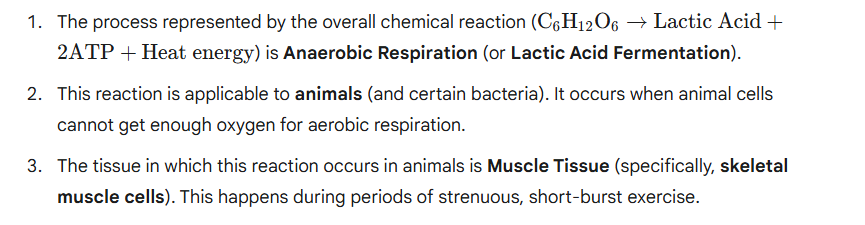
1.Given below is an overall chemical reaction of a certain process: C6H12O6 →LacticAcid+2ATP+Heat energy Name the process.
2. Given below is an overall chemical reaction of a certain process: C6H12O6→LacticAcid+2ATP+Heat energy Is this reaction applicable to animals or to plants or to both animals and plants?
3. Given below is an overall chemical reaction of a certain process: C6H12O6→LacticAcid+2ATP+Heat energy Name one tissue in which this reaction occurs.
Ans:
Question 3.
1. Given below are chemical reactions (1 to 5) involving glucose and five other chemical products (A-E).

Write the reaction number of the following:
(i) Anaerobic respiration in plants __________
(ii) End products in aerobic respiration ____________
(iii) Reaction occurring in liver _________
(iv) Anaerobic respiration in animals ________
(v) Storage in the liver _________
2. Given below are chemical reactions (1 to 5) involving glucose and five other chemical products (A-E).
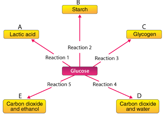
Which reactions (1-5) in the above correspond to the following (write the corresponding number of reactions next to them).
(i) Aerobic respiration
(ii) Change taking place in the liver
(iii) Anaerobic respiration in yeast
(iv) Change taking place in a plant storage organ, e.g., potato
(v) Anaerobic respiration in animals 3.
Ans:
1. Matching Reactions to Processes (Part 1)
| Process | Reaction Number |
| (i) Anaerobic respiration in plants | Reaction 5 (Produces CO2 and ethanol – characteristic of yeast/plants) |
| (ii) End products in aerobic respiration | Reaction 4 (Produces CO2 and water) |
| (iii) Reaction occurring in liver | Reaction 3 (Formation of Glycogen) |
| (iv) Anaerobic respiration in animals | Reaction 1 (Produces Lactic acid) |
| (v) Storage in the liver | Reaction 3 (Glucose is stored as Glycogen) |
2. Matching Reactions to Processes (Part 2)
| Process | Corresponding Reaction Number |
| (i) Aerobic respiration | Reaction 4 (Glucose breakdown into CO2 and H2O in the presence of O2) |
| (ii) Change taking place in the liver | Reaction 3 (Conversion of Glucose into Glycogen for storage) |
| (iii) Anaerobic respiration in yeast | Reaction 5 (Produces CO2 and ethanol) |
| (iv) Change taking place in a plant storage organ, e.g., potato | Reaction 2 (Conversion of Glucose into Starch for storage) |
| (v) Anaerobic respiration in animals | Reaction 1 (Produces Lactic acid) |
Question 4.
1. The volume of air in the lungs and the rate at which it is exchanged during inspiration and expiration was measured.The following diagram shows a group of lung volumes and capacities.

Study the diagram carefully and explain briefly the following : Tidal volume (TV)
2. The volume of air in the lungs and the rate at which it is exchanged during inspiration and expiration was measured.
The following diagram shows a group of the lung volumes and capacities.
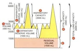
Study the diagram carefully and explain briefly the following : Inspiratory reserve volume (IRV)
3. The volume of air in the lungs and the rate at which it is exchanged during inspiration and expiration was measured.
The following diagram shows a group of the lung volumes and capacities.

Study the diagram carefully and explain briefly the following : Expiratory reserve volume (ERV)
4. The volume of air in the lungs and the rate at which it is exchanged during inspiration and expiration was measured.
The following diagram shows a group of the lung volumes and capacities.

Study the diagram carefully and explain briefly the following : Vital capacity (VC)
5. The volume of air in the lungs and the rate at which it is exchanged during inspiration and expiration was measured.
The following diagram shows a group of the lung volumes and capacities.

Study the diagram carefully and explain briefly the following : Residual volume (RV)
Ans:
The provided diagram illustrates various volumes and capacities of air exchanged in the lungs. Here are brief explanations for each:
1. Tidal Volume (TV)
The Tidal Volume is the volume of air that is inhaled or exhaled during a single, normal, quiet breathing cycle (approximately 500mL). It represents the amount of air exchanged when you are at rest.
2. Inspiratory Reserve Volume (IRV)
The Inspiratory Reserve Volume is the maximum volume of air that can be inhaled forcibly after a normal inspiration. It represents the extra air you can take in above the tidal volume (approximately 3000mL).
3. Expiratory Reserve Volume (ERV)
The Expiratory Reserve Volume is the maximum volume of air that can be exhaled forcibly after a normal expiration. It represents the extra air you can push out after a typical breath out (approximately 1000mL).
4. Vital Capacity (VC)
The Vital Capacity is the maximum volume of air a person can exchange in a single, maximal breath. It is the total amount of air that can be exhaled after a maximal inhalation. It is the sum of the Tidal Volume, Inspiratory Reserve Volume, and Expiratory Reserve Volume (VC = TV + IRV + ERV), approximately 4500mL.
5. Residual Volume (RV)
The Residual Volume is the volume of air that remains in the lungs and airways even after a maximal, forceful exhalation. This air cannot be expelled and ensures the lungs and alveoli do not completely collapse (approximately 1500 mL).
