
")
The chapter on Tissues begins by establishing the fundamental concept that a tissue is a group of cells that are similar in structure and work together to perform a specific function. In multicellular organisms, this division of labour is essential for efficiency. The chapter then classifies plant and animal tissues into broad categories. Plant tissues are first divided into Meristematic and Permanent tissues. Meristematic tissue consists of actively dividing cells found in growing regions like root and shoot tips, responsible for the plant’s increase in length. Once these cells lose their ability to divide and take on a specific role, they become Permanent tissue, which can be simple (composed of one cell type) or complex (composed of more than one cell type).
In plants, the study of permanent tissues covers several key types. Simple permanent tissues include Parenchyma (for storage and support), Collenchyma (for flexible support in young stems), and Sclerenchyma (for rigid support, with dead cells). Complex permanent tissues are the vascular tissues, which act as the plant’s transport system. Xylem carries water and minerals from roots to other parts, while Phloem transports the food prepared in the leaves to different areas for storage or use. Additionally, the chapter discusses the protective epidermal tissues that cover the plant and the cork that forms the bark in older stems and roots.
Shifting focus to animal tissues, the chapter explains that they are categorized based on their function and are more specialized. The four primary types are Epithelial tissue, which forms a protective covering on organs and the body; Connective tissue, which connects, supports, or separates other tissues (examples include bone, cartilage, blood, and ligaments); Muscular tissue, composed of cells that can contract and relax to enable movement; and Nervous tissue, which contains highly specialized neurons that make up the brain, spinal cord, and nerves, allowing for the control, coordination, and rapid response to stimuli. The chapter concludes by highlighting how the coordinated activity of these diverse tissues is what allows complex organisms to function as a single, integrated unit.
- Multiple choice type
Question 1.
In potato starch is stored in :
- Sclerenchyma
- Collenchyma
- Parenchyma
- Chlorenchyma
Question 2.
Tendons and ligaments are examples of
- Fibrous connective tissue
- Cartilage
- Muscular tissue
- Adipose tissue
Question 3.
Which one of the following pairs is correctly matched?
- Meristem – Actively dividing cells
- Xylem – Transport of food
- Phloem – Transport of water
- Sclerenchyma – Storage of starch
Question 4.
Parenchyma containing chloroplasts is known as :
- Parenchyma
- Aerenchyma
- Collenchyma
- Chlorenchyma
Question 5.
Annual rings are the number of ______.
- internodes in a stem
- rings of vascular bundles in a monocot stem
- barks layers in a woody stem
- layers of Xylem in a stem
Question 6.
Which of the following cells in plants are said to be nonliving?
- Meristem
- Parenchyma
- Collenchyma
- Sclerenchyma
Question 7.
Which of the following connects a muscle to a bone?
- Cartilage
- Ligament
- Tendon
- Interstitial fluid
Question 8.
Cardiac muscle is ______.
- Involuntary
- Smooth
- Striated
- Involuntary and Striated
- Very short answer type
Question 1.
1. Name the kind of tissue found at the tip of plant roots.
2. Name the kind of tissue found at the lower surface of the leaf.
3. Name the kind of tissue found in the inner lining of the intestine.
4. Name the kind of tissue found at the joint between two long bones.
5. Name the kind of tissue found in the walls of the veins of the leaves.
6. Name the kind of tissue found as gritty masses in the pulp of pears.
Ans:
Here are the names of the tissues found at the specified locations:
- Tip of plant roots: Apical Meristem (specifically the root apical meristem)
- Lower surface of the leaf: Epidermis (containing stomata and often guard cells)
- Inner lining of the intestine: Columnar Epithelium (specifically Simple Columnar Epithelium)
- Joint between two long bones: Cartilage (often hyaline cartilage) and Ligament (which connects bone to bone)
- Walls of the veins of the leaves: Vascular Tissue (composed of Xylem for water transport and Phloem for food transport)
- Gritty masses in the pulp of pears: Sclerenchyma (specifically Sclereids or Stone cells)
Question 2.
Where is the least specialized tissue located in plants?
Ans:
Here are the unique and concise descriptions for the biological terms you provided:
- A group of similar cells performing a specific function:Tissue
- Unique Description: An aggregate of biological cells that exhibit a similar structure and are unified to carry out a particular, collective task within an organism.
- Cells least specialized in plants:Parenchyma
- Unique Description: The most fundamental and undifferentiated type of plant cell, typically retaining the capacity to divide and responsible for basic functions like storage, healing, and photosynthesis.
- Cells responsible for the increase in diameter of the stem and root of dicot plants: Cambium (specifically the Vascular Cambium and Cork Cambium, which are forms of lateral meristem).
- Unique Description: The lateral meristematic layers that facilitate secondary growth, resulting in the widening (girth increase) of a dicot plant’s stem and root structure.
Question 3.
1. Write one word for the following: A group of similar cells performing a specific function.
2. Write one word for the following: Cells least specialized in plants.
3. Write one word for the following: Cells responsible for the increase in diameter of the stem and root of dicot plants.
Ans:
- A group of similar cells performing a specific function: Tissue
- Cells least specialized in plants: Parenchyma
- Cells responsible for the increase in diameter of the stem and root of dicot plants: Cambium (specifically the Vascular Cambium and Cork Cambium, which are forms of lateral meristem).
Question 4.
1. Name one place in living organisms where the following tissue is located: Meristematic tissue
2. Name one place in living organisms where the following tissues are located: Cartilage
3. Name one place in living organisms where the following tissues is located:
Squamous epithelium
4. Name one place in a living organism where the following tissues are located:
Sclerenchyma
5. Name one place in living organisms where the following tissue is located:
Ciliated epithelium
6. Name one place in living organisms where the following tissue is located:
Ligament
Ans:
| Tissue Type | Location (Primary Sites) | Functional Context |
| Meristematic Tissue | At the growing points, such as the apices of roots and stems (apical meristem). | Enables primary growth (increase in length) through continuous, rapid cell division. |
| Cartilage | Forms the flexible framework of the external ear (pinna) and the tip of the nose. Also caps the ends of long bones within movable joints. | Provides flexible support and acts as a shock absorber between articulating bones. |
| Squamous Epithelium | Lines delicate surfaces, such as the inside of the cheek (buccal cavity) and the thin layer covering the air sacs (alveoli) of the lungs. | Forms a smooth, minimally obstructive boundary ideal for diffusion (like gas exchange). |
| Sclerenchyma | Found in parts requiring maximum rigidity, such as the husk of coconut and the gritty texture (stone cells) in the pulp of pears. | Offers robust mechanical support and protection against compression and shear forces. |
| Ciliated Epithelium | Covers the interior surface of the trachea (windpipe) and the larger respiratory tubes (bronchi). | Uses synchronized beating of hair-like cilia to sweep mucus and filtered debris out of the respiratory tract. |
| Ligament | Located at skeletal joints, where it forms a band that connects one bone to another bone (e.g., knee or ankle joints). | Provides joint stability while allowing controlled, limited movement. |
Question 5.
1. Name the kind of cells found in the following place: Surface of the human skin
2. Name the kind of cell found in the following place: Salivary gland
3. Name the kind of cells found in the following place: Brain
4. Name the kind of cells found in the following place: Inner lining of the wind pipe
Ans:
Here are the kinds of cells found in the specified locations:
- Surface of the human skin: Squamous Epithelial Cells (specifically forming the stratified squamous epithelium for protection).
- Salivary gland: Glandular Epithelial Cells (specifically forming the secretory units or acini) and Cuboidal Epithelial Cells (lining the ducts).
- Brain: Neurons (Nerve cells, responsible for transmitting signals) and various types of Neuroglia (Glial cells, which support and protect neurons).
- Inner lining of the windpipe (Trachea): Ciliated Columnar Epithelial Cells (which have hair-like cilia to sweep mucus and dust away) and Goblet cells (which secrete mucus).
- Short answer type
Question 1.
Name any one body part where ciliated epithelium is found in humans. What is its function?
Ans:
Ciliated epithelium is found in the lining of the human trachea (windpipe) and the larger bronchi of the respiratory tract.
Function of Ciliated Epithelium
- Mucus Trapping: Glandular cells within the epithelium produce mucus that traps inhaled dust particles, pathogens (bacteria, viruses), and other foreign matter.
- Ciliary Movement: The cilia (hair-like projections) on the cell surface beat in a coordinated, rhythmic, wave-like motion.
- Sweeping Action: This continuous beating sweeps the debris-laden mucus upward, away from the lungs, toward the pharynx (throat), where it can be swallowed or coughed out. This action keeps the lower respiratory passages clean and protects the lungs from infection and damage.
Question 2.
What is the difference between the nervous tissue and the nervous system?
Ans:
The difference between nervous tissue and the nervous system lies in their level of organization and scope: nervous tissue is the fundamental material (cellular level), while the nervous system is the entire functional and structural network (organ system level).
Nervous Tissue
Nervous tissue is the basic biological material that makes up the nervous system.
- Definition: It is one of the four principal types of tissue in the body, composed primarily of specialized cells: neurons (which transmit electrical signals) and neuroglia (or glial cells, which provide support and insulation).
- Function: Its function is to generate, transmit, and process electrical and chemical signals.
- Location: Found everywhere the nervous system exists, forming the brain, spinal cord, and nerves.
Nervous System
The nervous system is the complete, organized network of organs and structures in the body built from nervous tissue.
- Definition: It is the body’s master control and communication system that coordinates all actions and functions.
- Components: It includes the Central Nervous System (CNS) (brain and spinal cord) and the Peripheral Nervous System (PNS) (all the nerves extending from the CNS).
- Function: Its function is to rapidly receive sensory information, interpret it, and initiate responses in other body parts (like muscles and glands).
Question 3.
List the tissues found in the human heart.
Ans:
Here are the main tissues found in the human heart:
Tissues in the Human Heart
1. Muscular Tissue (The Pumping Engine)
- Cardiac Muscle Tissue: This is the most crucial tissue, forming the thick walls (myocardium) of the heart. It is characterized by striated, involuntary, and branched fibers connected by intercalated discs. Its primary function is rhythmic contraction to pump blood.
2. Connective Tissue (Support and Structure)
- Fibrous Connective Tissue: Provides the structural framework, forming the heart’s valves, the septa (walls dividing chambers), and the outer fibrous layer (pericardium).
- Areolar Connective Tissue: Fills the spaces and holds the muscle fibers together.
- Blood: While fluid, blood is considered a specialized connective tissue that flows through the heart chambers.
3. Epithelial Tissue (Lining and Protection)
- Endothelium: This simple squamous epithelium forms the smooth, innermost lining of the heart chambers and blood vessels, known as the endocardium. This lining ensures friction-free blood flow.
- Mesothelium: A type of simple squamous epithelium that forms the serous layer of the pericardium (the sac around the heart).
4. Nervous Tissue (Regulation)
- Nerves: The heart contains nervous tissue (nerve fibers) that originate from the autonomic nervous system. These nerves regulate the rate and force of heart contractions (though the heart itself has its own pacemaker system, the nervous tissue modulates it).
Question 4.
Can you consider a cluster of eggs as a tissue? Why?
Ans:
Why a Cluster of Eggs is Not a Tissue
The definition of a tissue is based on organization, function, and structure. A cluster of eggs fails to meet this definition for the following reasons:
- Lack of Organization and Integration: A tissue is defined as a group of similar cells (or sometimes dissimilar cells with a common origin) that are structurally organized and integrated to perform a specific common function. Individual eggs, or ova, are single, highly specialized reproductive cells that function independently of one another. They are not joined by the cell junctions characteristic of tissues.
- Difference in Cellular Function: Eggs are gametes (sex cells) whose sole biological purpose is reproduction (fusion with a sperm). A cluster simply represents a quantity of these individual reproductive cells grouped together, often by a separate surrounding structure (like an egg case or follicle). They do not collectively perform a common function like contraction (muscle tissue) or protection (epithelial tissue).
- Level of Organization: Biological organization progresses from cells to tissues, organs, and organ systems.
- Eggs are at the cellular level.
- Tissues are at the supra-cellular level.
Question 5.
Name the three kinds of muscles found in the human body. In each case, name one region in the body where they are found.
Ans:
| Muscle Type | Characteristics | Region Found |
| 1. Skeletal Muscle (Striated/Voluntary) | Striated (banded), cylindrical fibers, multinucleated, under voluntary control. | Attached to bones (e.g., muscles of the arms, legs, or back). |
| 2. Smooth Muscle (Non-striated/Involuntary) | Non-striated, spindle-shaped fibers, single nucleus, under involuntary control. | Walls of hollow internal organs (e.g., stomach, intestines, blood vessels, urinary bladder). |
| 3. Cardiac Muscle (Striated/Involuntary) | Striated, branched fibers, single nucleus, connected by intercalated discs, under involuntary control. | Walls of the Heart (myocardium). |
- Long answer type
Question 1.
1. What is the difference between cells and tissue?
2. What is the difference between an organ and an organism?
3. What is the difference between organ and organelle?
4. What is the difference between organ and organ system?
Ans:
1. Cells and Tissue
| Feature | Cell | Tissue |
| Level | Basic structural and functional unit of life. | A group of similar cells working together. |
| Organization | The simplest unit of life; contains organelles. | A collection of cells organized and integrated to perform a specific common function (e.g., muscle, nervous, epithelial). |
| Example | A muscle fiber, a nerve cell (neuron), an epithelial cell. | Muscle tissue, nervous tissue, epithelial tissue. |
2. Organ and Organism
| Feature | Organ | Organism |
| Level | A specialized structure composed of different tissues. | A complete, individual living entity. |
| Composition | Made up of multiple tissues (e.g., muscle, nervous, and connective) working together. | Made up of multiple organ systems (e.g., digestive, circulatory, nervous) working interdependently. |
| Example | The heart, the stomach, the liver. | A human being, a tree, a dog, a bacterium. |
3. Organ and Organelle
| Feature | Organ | Organelle |
| Level | A large structure composed of tissues (supracellular level). | A small, specialized structure found inside a cell (subcellular level). |
| Size/Visibility | Large; macroscopic (visible to the naked eye). | Microscopic; visible only with an electron microscope. |
| Function | Performs a major body function (e.g., digestion, pumping blood). | Performs a specific function within the cell (e.g., respiration, protein synthesis). |
| Example | The lung, the kidney. | Mitochondrion, Ribosome, Nucleus. |
4. Organ and Organ System
| Feature | Organ | Organ System |
| Level | A structure composed of different tissues. | A group of different organs working together. |
| Function | Performs a single, specific major function (e.g., the stomach performs the mixing and initial digestion of food). | Performs a complex series of integrated functions necessary for survival (e.g., the digestive system handles all aspects of food breakdown and absorption). |
| Components | Contains tissues (e.g., stomach contains muscle and epithelial tissue). | Contains organs (e.g., digestive system contains the stomach, small intestine, and liver). |
Question 2.
1. Differentiate between cells of Parenchyma and Collenchyma.
2. Differentiate between cells of Meristematic tissue and Permanent tissue
3. Differentiate between cells of Sclerenchyma and Parenchyma
4. Differentiate between cells of cells of involuntary and voluntary muscle
5. Differentiate between cells of Fibres of voluntary muscle and cardiac muscle
Ans:
1. Parenchyma and Collenchyma
| Feature | Parenchyma Cells | Collenchyma Cells |
| Specialization | Least specialized; generally isodiametric (spherical/oval). | Moderately specialized; often elongated. |
| Cell Wall | Thin and uniform primary cell walls. | Cell walls are unevenly thickened, especially at the corners, with cellulose and pectin. |
| Nature | Usually living cells at maturity. | Living cells at maturity. |
| Function | Storage, photosynthesis, gas exchange, packing, and healing. | Provides mechanical support and flexibility (plastic support) to young stems and petioles. |
| Location | Cortex, pith, mesophyll, and storage organs. | Beneath the epidermis in young stems and leaf stalks. |
2. Meristematic tissue and Permanent tissue
| Feature | Meristematic Tissue | Permanent Tissue |
| Cell Division | Cells are actively and continuously dividing (mitotic). | Cells have lost the power to divide (or divide only under specific conditions). |
| Cell Status | Cells are undifferentiated (immature), helping in plant growth. | Cells are fully differentiated (mature), helping in protection, conduction, or support. |
| Metabolism | High metabolic rate. | Low metabolic rate. |
| Vacuoles | Small or absent. | Often have large central vacuoles (especially parenchyma). |
3. Sclerenchyma and Parenchyma
| Feature | Sclerenchyma Cells | Parenchyma Cells |
| Cell Wall | Cell walls are thickened uniformly with lignin, making them hard and rigid. | Cell walls are thin and composed only of cellulose. |
| Nature | Typically dead (non-living) at maturity. | Typically living at maturity. |
| Function | Provides mechanical strength and structural rigidity (elastic support). | Storage, photosynthesis, and gas exchange. |
| Types | Fibers (long, pointed) and Sclereids (stone cells, gritty). | Isodiametric, thin-walled. |
4. cells of involuntary and voluntary muscle
| Feature | Voluntary Muscle Cells (Skeletal Muscle) | Involuntary Muscle Cells (Smooth Muscle) |
| Control | Contraction is under conscious, willful control (somatic nervous system). | Contraction is not under conscious control (autonomic nervous system). |
| Striations | Striated (show light and dark bands/stripes). | Non-striated (smooth or unstriped appearance). |
| Nucleus | Multinucleated (syncytial), with nuclei located at the periphery. | Uninucleated, with the nucleus centrally located. |
| Location | Attached to bones (skeleton) for locomotion. | Walls of internal hollow organs (e.g., stomach, intestine, blood vessels). |
5. Fibres of voluntary muscle and cardiac muscle
| Feature | Voluntary Muscle Fibers (Skeletal) | Cardiac Muscle Fibers |
| Control | Voluntary (under conscious control). | Involuntary (not under conscious control). |
| Branching | Unbranched, cylindrical fibers. | Branched fibers, forming an interconnected network. |
| Intercalated Discs | Absent. | Present. These junctions allow rapid communication, ensuring synchronized contraction. |
| Nucleus | Multiple nuclei located peripherally. | Usually one or two nuclei located centrally. |
| Function | Body movement and posture. | Rhythmic, continuous pumping of blood. |
- Structured/Application Questions
Question 1.
1.Study the diagram given below and then answer the question that follows:

Identify the tissue and give a reason to support your answer.
2. Study the diagram given below and then answer the question that follows:
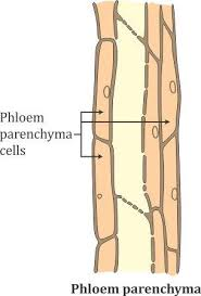
Name the parts labelled 1, 2, 3 and 4.
3. Study the diagram given below and then answer the question that follows:

Where is this tissue likely to be found in the plant?
4. Study the diagram given below and then answer the question that follows:

State the function of the parts labelled 1, 2, 3 and 4.
Ans:
1. Identify the Tissue and Give a Reason
The tissue shown in the diagram is Collenchyma tissue.
Reason: The defining feature is the uneven thickening of the cell walls (visible, especially where cells meet), while the cells themselves remain living (indicated by the presence of a nucleus/cytoplasm) and elongated. This structure provides flexible support without restricting growth.
2. Name the Parts Labelled 1, 2, 3 and 4
The labels point to the structural components of the collenchyma cells:
- 1: Unevenly Thickened Cell Wall
- 2: Cytoplasm
- 3: Nucleus
- 4: Parenchyma Cells (These are typically adjacent to or surrounding the Collenchyma tissue, serving as the ground tissue).
3. Where is This Tissue Likely to be Found?
Collenchyma tissue is found in areas of the plant that require flexible mechanical support and are still growing.
It is typically located:
- Beneath the epidermis of young stems (especially herbaceous dicot stems).
- In the petioles (leaf stalks) and the main veins of leaves.
4. State the Function of the Parts Labelled 1, 2, 3 and 4
The functions correspond to the labeled structures:
- 1 (Unevenly Thickened Cell Wall): Provides mechanical support and tensile strength to the plant parts without making them rigid, allowing them to bend without breaking.
- 2 (Cytoplasm): The site of metabolic activities and holds the cell’s organelles. Since collenchyma cells are living, the cytoplasm is active.
- 3 (Nucleus): Controls all cellular activities, including cell division (under specific circumstances) and the synthesis of materials.
- 4 (Parenchyma Cells): Functions primarily in storage of food and water, or, if they contain chloroplasts, in photosynthesis (as chlorenchyma).
Question 2.
1. Study the diagram given below and then answer the question that follows:

Identify the cell.
2. Study the diagram given below and then answer the question that follows:
Name the parts labelled 1, 2, 3, 4, 5 and 6.
3. Study the diagram given below and then answer the questions that follow:
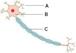
Where is this cell likely to be found in the human body and what is its function?
Ans:
1. Identify the Cell
The cell shown in the diagram is a Neuron or Nerve Cell.
2. Name the Parts Labelled 1, 2, 3, 4, 5 and 6
The labels point to the structural components of the neuron:
- 1: Dendrites (or Dendrons)
- 2: Axon
- 3: Nucleus
- 4: Cyton (or Cell Body / Soma)
- 5: Myelin Sheath (The insulating layer surrounding the axon)
- 6: Nerve Endings (or Axon Terminals)
3. Location and Function of the Cell
Location:
The neuron is the primary cell type of the Nervous System. It is found throughout the human body, forming:
- The Brain and Spinal Cord (Central Nervous System).
- The Nerves that extend to all parts of the body (Peripheral Nervous System).
Function:
The primary function of the neuron is conduction of information. Specifically:
- To receive stimuli (via dendrites).
- To transmit rapid electrical and chemical signals (impulses) over long distances (via the axon) to coordinate all body activities, thoughts, and senses.
