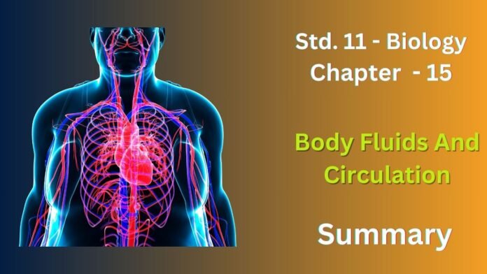
Body fluids are essential for the transport of nutrients, oxygen, waste products, and hormones throughout the body. The circulatory system plays a crucial role in the circulation of these fluids.
Blood
- Composition: Blood is a connective tissue composed of plasma (liquid component) and cells (red blood cells, white blood cells, and platelets).
- Functions:
- Transportation of oxygen, carbon dioxide, nutrients, hormones, and waste products.
- Immunity and defense against pathogens.
- Clotting to prevent blood loss.
Blood Vessels
- Arteries: Carry blood away from the heart.
- Veins: Carry blood back to the heart.
- Capillaries: Tiny vessels that allow for the exchange of substances between blood and tissues.
The Heart
- Structure: A muscular organ divided into four chambers (right atrium, right ventricle, left atrium, left ventricle).
- Function: Pumps blood throughout the body.
Circulatory Pathways
- Systemic Circulation: Blood flows from the heart to the body tissues and back to the heart.
Lymphatic System
- Function: Drains excess fluid from tissues, filters lymph, and helps in immunity.
- Lymph Nodes: Filter lymph and contain white blood cells.
Disorders of the Circulatory System
- Atherosclerosis: Buildup of plaque in arteries, leading to heart disease.
- Hypertension: High blood pressure.
- Anemia: Deficiency of red blood cells or hemoglobin.
- Leukemia: Cancer of blood-forming cells.
Exercise
1. Name the components of the formed elements in the blood and mention one major function of each of them
Ans :
Formed elements are the cellular components of blood. They make up about 45% of the total blood volume.
- Red Blood Cells (Erythrocytes):
- Function: Transport oxygen from the lungs to tissues and carbon dioxide from tissues to the lungs.
- White Blood Cells (Leukocytes):
- Function: Fight infection and protect the body from foreign invaders.
- Platelets (Thrombocytes):
- Function: Help in blood clotting to prevent blood loss.
2. What is the importance of plasma proteins?
Ans :
Plasma proteins are essential components of blood plasma, making up about 7% of its total volume. They play a variety of crucial roles in the body, including:
- Maintaining osmotic pressure: Albumin, the most abundant plasma protein, helps to maintain the osmotic pressure of the blood, which is important for regulating blood volume and preventing fluid from leaking into tissues.
- Transport: Many plasma proteins act as carriers, transporting various substances throughout the body. For example, albumin transports fatty acids, hormones, and certain drugs. Globulins transport iron, copper, and other substances.
- Clotting: Several plasma proteins, including fibrinogen and prothrombin, are involved in the blood clotting process. These proteins help to form a clot to prevent blood loss after an injury.
- Immunity: Antibodies, which are a type of protein, are essential for the immune system to fight off infections.
- Nutrient transport: Some plasma proteins, such as transferrin, transport nutrients like iron.
3. Match Column I with Column II.
Column I Column II
(a) Eosinophils (i) Coagulation
(b) RBC (ii) Universal recipient
(c) AB Group (iii) Resist infections
(d) Platelets (iv) Contraction of heart
(e) Systol (v) Gas transport
Ans :
(a) Eosinophils – (iii) Resist infections
(b) RBC – (v) Gas transport
(c) AB Group – (ii) Universal recipient
(d) Platelets – (i) Coagulation
(e) Systol – (iv) Contraction of heart
4. Why do we consider blood as a connective tissue?
Ans :
Blood is considered a connective tissue because it shares several key characteristics with other connective tissues:
- Extracellular Matrix: Blood is composed of a liquid matrix (plasma) and various cellular components (formed elements). This extracellular matrix provides a medium for the transport of substances throughout the body.
- Connects Tissues: Blood connects different tissues and organs by transporting nutrients, oxygen, waste products, and hormones.
- Support and Protection: Blood plays a crucial role in supporting and protecting the body by maintaining homeostasis, transporting oxygen and nutrients, and fighting infections.
- Derived from Mesoderm: Like other connective tissues, blood is derived from the mesoderm, one of the three primary germ layers during embryonic development.
5. What is the difference between lymph and blood?
Ans :
| Feature | Blood | Lymph |
| Color | Red | Colorless or slightly yellowish |
| Composition | Red blood cells, white blood cells, platelets, plasma | White blood cells, lymph fluid |
| Circulatory System | Cardiovascular system | Lymphatic system |
| Functions | Transports oxygen, carbon dioxide, nutrients, hormones, waste products; immunity, clotting | Drains excess fluid, transports white blood cells, absorbs fats |
| Oxygen Level | Higher | Lower |
| Protein Content | Higher | Lower |
| Flow Rate | Faster | Slower |
| Clotting | Clots more quickly | Does not clot as easily |
6. What is meant by double circulation? What is its significance?
Ans :
Double Circulation refers to the circulatory system in vertebrates where blood passes through the heart twice in a complete circuit. This system is more efficient than single circulation, allowing for better oxygenation of tissues and removal of waste products.
Significance of Double Circulation:
- Efficient Oxygenation: Blood passes through the lungs for oxygenation before being pumped to the body’s tissues, ensuring that tissues receive a sufficient supply of oxygen.
- Waste Removal: Deoxygenated blood returns to the heart for purification before being pumped to the lungs to eliminate carbon dioxide and other waste products.
- Separation of Oxygenated and Deoxygenated Blood: Double circulation maintains a relatively separate flow of oxygenated and deoxygenated blood, preventing mixing and ensuring efficient oxygen delivery.
- Higher Metabolic Rates: The efficiency of double circulation allows for higher metabolic rates and supports more complex organisms.
7. Write the differences between :
(a) Blood and Lymph
(b) Open and Closed system of circulation
(c) Systole and Diastole
(d) P-wave and T-wave
Ans :
(a) Blood and Lymph
| Feature | Blood | Lymph |
| Color | Red | Colorless or slightly yellowish |
| Composition | Red blood cells, white blood cells, platelets, plasma | White blood cells, lymph fluid |
| Circulatory System | Cardiovascular system | Lymphatic system |
| Functions | Transports oxygen, carbon dioxide, nutrients, hormones, waste products; immunity, clotting | Drains excess fluid, transports white blood cells, absorbs fats |
| Oxygen Level | Higher | Lower |
| Protein Content | Higher | Lower |
| Flow Rate | Faster | Slower |
| Clotting | Clots more quickly | Does not clot as easily |
(b) Open and Closed system of circulation
| Feature | Open System | Closed System |
| Circulatory Fluid | Hemolymph | Blood |
| Vessels | Open-ended vessels (sinuses) | Closed network of arteries, veins, and capillaries |
| Direct Contact | Hemolymph directly bathes tissues | Blood does not directly contact tissues |
| Efficiency | Less efficient, slower circulation | More efficient, rapid circulation |
| Examples | Insects, crustaceans | Vertebrates, earthworms |
(c) Systole and Diastole
| Feature | Systole | Diastole |
| Phase | Contraction of the heart muscle | Relaxation of the heart muscle |
| Chambers Involved | Atria and ventricles contract | Atria and ventricles relax |
| Blood Flow | Blood is pumped out of the heart | Blood fills the heart chambers |
| Pressure | Blood pressure rises | Blood pressure falls |
(d) P-wave and T-wave
| Feature | P-wave | T-wave |
| Represents | Atrial depolarization (contraction) | Ventricular repolarization (relaxation) |
| Shape | Rounded, upward peak | Rounded, downward peak |
| Timing | Before the QRS complex | After the QRS complex |
| Significance | Indicates electrical activity in the atria | Indicates the end of ventricular contraction and the beginning of ventricular relaxation |
8. Describe the evolutionary change in the pattern of heart among the vertebrates.
Ans :
Vertebrates have evolved a complex circulatory system that has undergone significant changes throughout their evolutionary history. Here’s a brief overview of these changes:
- Single-loop Circulation: In fish, the circulatory system is a single-loop system. Blood is pumped from the heart to the gills for oxygenation and then flows to the rest of the body before returning to the heart. This system is less efficient in delivering oxygen to tissues.
- Double-loop Circulation: Amphibians, reptiles, birds, and mammals have a double-loop circulatory system. This system separates oxygenated and deoxygenated blood, allowing for more efficient oxygen delivery to tissues.
- Pulmonary Circulation: Blood is pumped from the heart to the lungs for oxygenation and then returns to the heart.
- Systemic Circulation: Oxygenated blood is pumped from the heart to the body tissues and then returns to the heart, where it is deoxygenated.
- Three-chambered Heart: Amphibians have a three-chambered heart, with two atria and a single ventricle. This allows for some mixing of oxygenated and deoxygenated blood, reducing the efficiency of oxygen delivery.
- Four-chambered Heart: Reptiles, birds, and mammals have a four-chambered heart, with two atria and two ventricles. This complete separation of oxygenated and deoxygenated blood ensures efficient oxygen delivery to tissues.
9. Why do we call our heart myogenic?
Ans :
Our hearts are myogenic because they generate their own electrical impulses. This means that the heart muscle itself is capable of initiating and regulating its own contractions, without the need for external signals from the nervous system.
This property is due to the presence of specialized cells called autorhythmic cells within the heart muscle. These cells have the ability to spontaneously depolarize and generate action potentials, which spread throughout the heart muscle and trigger contractions.
This intrinsic ability of the heart to generate its own electrical impulses is essential for maintaining a regular heartbeat and ensuring adequate blood flow to the body. It allows the heart to function independently, even in the absence of external innervation.
10. Sino-atrial node is called the pacemaker of our heart. Why?
Ans :
The sino-atrial (SA) node is often referred to as the “pacemaker of the heart” because it is the primary source of electrical impulses that initiate the heartbeat.
Located in the right atrium of the heart, the SA node is a specialized group of cardiac muscle cells that have the ability to spontaneously generate electrical signals. These signals spread throughout the heart muscle, causing it to contract and pump blood.
12. What is the significance of atrio-ventricular node and atrio-ventricular bundle in the functioning of heart?
Ans :
Atrio-ventricular (AV) node and atrio-ventricular bundle play crucial roles in the electrical conduction system of the heart, ensuring coordinated contractions of the atria and ventricles.
Atrio-ventricular (AV) node:
- Location: Located at the junction between the atria and ventricles.
- Function: Delays the electrical impulse from the atria to the ventricles, allowing the atria to contract and fill the ventricles with blood before the ventricles contract.
- Significance: This delay ensures that the ventricles have enough time to fill with blood before they pump it out of the heart.
Atrio-ventricular (AV) bundle:
- Location: Also known as the bundle of His, it extends from the AV node through the interventricular septum.
- Function: Conducts the electrical impulse from the AV node to the ventricles.
- Significance: This ensures that the electrical impulse reaches all parts of the ventricles, causing them to contract simultaneously and pump blood effectively.
13. Define a cardiac cycle and the cardiac output.
Ans :
Cardiac Cycle
It involves the contraction and relaxation of the heart’s chambers to pump blood throughout the body.
Cardiac Output
It is calculated by multiplying the heart rate (number of beats per minute) by the stroke volume (volume of blood pumped per beat).
14. Explain heart sounds
Ans :
Heart sounds are the noises produced by the heart’s valves closing and the contraction of the heart muscle. These sounds can be heard using a stethoscope and are used to assess the health of the heart.
There are two main heart sounds:
- S1 (First Heart Sound):
- Cause: Closure of the atrioventricular (AV) valves (mitral and tricuspid valves).
- Timing: Occurs at the beginning of ventricular systole.
- Sound: A dull, low-pitched “lub” sound.
- S2 (Second Heart Sound):
- Cause: Closure of the semilunar valves (aortic and pulmonary valves).
- Sound: A sharper, higher-pitched “dub” sound.
15. Draw a standard ECG and explain the different segments in it.
Ans :
- P wave: Indicates atrial depolarization, the electrical activity that causes the atria to contract.
- T wave: Reflects ventricular repolarization, the electrical activity that leads to ventricular relaxation.
- PR interval: Measures the time from the start of the P wave to the start of the QRS complex, representing the duration it takes for the electrical impulse to travel from the atria to the ventricles.
- QRS interval: Indicates the duration of the QRS complex, representing the time required for the ventricles to depolarize and contract.
- ST segment: The flat segment between the QRS complex and the T wave, representing the interval between ventricular depolarization and repolarization.
- QT interval: Measures the time from the beginning of the QRS complex to the end of the T wave, representing the total time needed for the ventricles to depolarize and repolarize.


