
 (1280 x 720 px) (25)")
The cell’s journey, from birth to division, is called the cell cycle, comprising two main phases: Interphase, for growth and DNA replication, and M-phase, for actual division.
Two crucial types of cell division exist:
- Mitosis yields two genetically identical daughter cells, each with the same chromosome count as the parent. It’s vital for growth, tissue repair, and asexual reproduction, involving precise chromosome organization and separation through stages: prophase, metaphase, anaphase, and telophase.
- Meiosis, conversely, produces four unique daughter cells, each containing half the parent’s chromosome number. This two-step process (Meiosis I and Meiosis II) is essential for sexual reproduction and generates genetic diversity, notably through “crossing over.”
Our genetic blueprint is stored in chromosomes, thread-like structures in the nucleus. These are composed of DNA tightly wound around histone proteins. Key terms include chromatin (the DNA-protein complex), chromatids (identical DNA copies), the centromere (where sister chromatids connect), and telomeres (protective chromosome ends).
In essence, this chapter illuminates how cells propagate, meticulously safeguarding and transmitting genetic information, thereby contributing to life’s vast diversity.
A. MULTIPLE CHOICE TYPE (Choose the best option out of the four alternatives a, b, c, and d)
1) The Chromatin material is formed forms of
(a) DNA only
(b) DNA and histones
(c) Histones only
(d) Nucleotides
Ans: (b) DNA and Histones
2) The term “chromosomes” literally means
(a) Inherited bodies
(b) Twisted threads
(c) Coloured bodies
(d) Shining threads Solution
Ans: (c) Coloured bodies
B. VERY SHORT ANSWER TYPE:
1)Name the following:
a) The repeating components of each DNA strand lengthwise.
b) The complex consists of a DNA strand and a core of histones.
c) The type of bond which joins the complementary nitrogenous bases.
d) The three components of a nucleotide.
Ans: (a) Nucleotides.
(b) Nucleosome.
(c) Hydrogen Bond.
(d) Phosphate, Sugar and Nitrogenous base.
C. SHORT ANSWER TYPE
1) What is the difference between chromatin fibre and chromosome?
Ans: Chromatin is the loose, thread-like DNA-protein complex found in the nucleus during cell growth (interphase), enabling gene expression. A chromosome is the tightly packed form of chromatin, visible during cell division, which ensures precise distribution of genetic material to new cells.
2)What are the rungs of the “DNA ladder” made of? Solutions:
Ans:The “rungs” of the DNA ladder are formed by specific pairings of nitrogenous bases: Adenine (A) always binds with Thymine (T) via two hydrogen bonds, while Guanine (G) always pairs with Cytosine (C) using three hydrogen bonds. This exact pairing is fundamental to DNA’s function.
2) Correct the following statements if there is any mistake.
a) The four nitrogenous bases in the DNA are Guanine, Thiamine, Adrenaline and Cytosine. b) Genes are specific sequences of bases on a chromosome.
c) A nucleotide is composed of a sulphate, a sugar (pentose) and a nitrogenous base.
d) Nucleosomes are groups of cysteine molecules surrounded by DNA strands.
e) If there are 46 chromosomes in a cell there will be 23 chromatin fibres inside the nucleus during interphase.
Ans: (a) The four nitrogenous bases in the DNA ladder are Guanine, Thymine, Adenine and Cytosine.
(b) Genes are specific sequences of nucleotides on a chromosome.
(c) A nucleotide is composed of a phosphate, sugar (pentose) and a nitrogenous base.
(d) Nucleosomes are groups of histone molecules surrounded by DNA strands.
(e) If there are 46 chromosomes in a cell there will be 46 chromatin fibres inside the nucleus during interphase.
D. LONG ANSWER TYPE:
1) What is a nucleosome?
Ans: A nucleosome is chromatin’s basic repeating unit. It’s formed by DNA wrapped around a core of eight histone proteins, compacting DNA to fit within the cell nucleus.
2)What are genes?
Ans: Genes are DNA segments that hold instructions for an organism’s development and maintenance. As the fundamental units of heredity, they pass from parents to offspring, influencing traits such as eye color or disease susceptibility.
E. STRUCTURED / APPLICATION / SKILL TYPE:
1)Given below is a schematic diagram of a portion of DNA.
(a) How many strands are shown in the pic?
(b) How many nucleotides have been shown in each strand?
(c) Name the parts numbered 1, 2, 3, 4 and 5 respectively.
(d) Name the DNA unit constituted by the parts 1, 2, 3 collectively.

Ans:
(a) Observing the schematic diagram, we can clearly identify two distinct strands. These strands are shown running antiparallel to each other, a characteristic feature of DNA. Each strand is composed of a backbone made of alternating sugar (represented by the pentagon labeled ‘2’) and phosphate groups (represented by the circle labeled ‘1’). Extending inwards from each sugar are nitrogenous bases (represented by the rectangles ‘3’ and ‘5’). These bases pair up across the two strands (indicated by the connections labeled ‘4’), forming the “rungs” of the DNA ladder.
(b) To determine the number of nucleotides in each strand, we need to count the complete repeating units on both the upper and lower strands.
- Upper strand: On the top strand, we can observe two distinct units, each composed of a phosphate group (labeled as 1, represented by a circle), a deoxyribose sugar (labeled as 2, represented by a pentagon), and a nitrogenous base (represented by the rectangular shapes labeled 3 and 5). Therefore, there are two complete nucleotides shown on the upper strand.
- Lower strand: Similarly, on the bottom strand, we can identify two complete units, each consisting of a phosphate group, a deoxyribose sugar, and a nitrogenous base. Thus, there are also two complete nucleotides shown on the lower strand.
In summary, the diagram depicts two nucleotides on each strand, making a total of four nucleotides in this depicted portion of the DNA double helix.
(c) The parts are: 1 – Phosphate group, 2 – Deoxyribose sugar, 3 – Nitrogenous base (Purine, likely Adenine or Guanine), 4 – Hydrogen bonds, 5 – Nitrogenous base (Pyrimidine, likely Thymine or Cytosine).
(d) Parts 1, 2, and 3 collectively form a nucleotide.
2) The three sketches (A, B and C) are intended to represent the replication of DNA. What should be their correct sequence starting with the first and ending with the last? ……
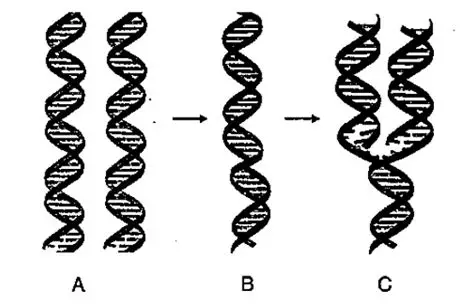
Ans: Stage A shows two separate, intact double-helix DNA molecules. This represents the state before replication has begun. Essentially, these are the parent DNA molecules in their original form.
Stage B depicts a single double-helix DNA molecule where the two strands are starting to unwind or separate. This is the crucial initial step of DNA replication, where the hydrogen bonds between the complementary base pairs break, allowing the strands to “unzip.” This separation creates a replication fork, making the individual strands available as templates.
Stage C illustrates the ongoing process of replication. Here, the original DNA molecule has partially unwound, and new complementary strands are being synthesized alongside each of the separated original strands. This results in two new DNA molecules, each composed of one original (parental) strand and one newly synthesized strand. This is characteristic of semi-conservative replication.
- A represents the starting point with complete DNA molecules.
- B shows the initial unwinding of a single DNA molecule.
- C illustrates the synthesis of new strands, leading to two nascent DNA molecules.
MULTIPLE CHOICE TYPE: (Choose the best option out of the four alternatives a, b, c and d)
1) The number of chromosomes in a certain type of cell division is halved. This kind of cell division occurs in.
(a) Only testis
(b) Only ovary
(c) Both ovary and testis
(d) All body cells
Ans: (c) Both ovary and testis
2) In which one of the following options the two stages of mitosis have been given in the correct sequence?
(a) Prophase, anaphase
(b) Metaphase, telophase
(c) Anaphase, telophase
(d) Telophase, anaphase
Ans: (c) Anaphase, telophase
3) Synthesis phase in the cell cycle is called so for the synthesis of more of.
(a) RNA
(b) RNA and proteins
(c) DNA
(d) Glucose
Ans: (c) DNA
B. VERY SHORT ANSWER TYPE:
1) Imagine one cell
(A) has undergone one mitotic division and another cell
(B) has completed its meiotic division. How many cells would the two produce?
Ans: A single mitotic division of Cell A will produce two genetically identical daughter cells. Mitosis is essential for growth, tissue repair, and asexual reproduction in somatic cells, ensuring each new cell receives a full, identical set of chromosomes.
Cell B ,This two-stage division (Meiosis I and Meiosis II) occurs in germline cells, creating genetically unique gametes. The reduction in chromosome number and genetic variation, largely due to crossing over and independent assortment, are key outcomes of meiosis.
2) Match the events given in column A with the phase in mitotic cell division in column B Column ‘A’ Column ‘B’
a) Chromosomes get arranged in a horizontal plane at the equator.
Ans: Prophase
b) Daughter chromosomes move to the opposite poles of a spindle.
Ans: Anaphase
c) Chromosomes become visible as fine long threads.
Ans: Metaphase
d) Chromosomes lose their distinctiveness and gradually become transformed into a chromatin network.
Ans:Telophase
3) Fill in the blanks:
a) Mitosis occurs in our ______ cells
Ans: (a) Somatic (body)
b) Mitosis produces two daughter cells whereas meiosis produces ______ daughter cells.
Ans: (b) Four
c) Meiosis occurs only in __________ cells.
Ans: (c) Reproductive
d) Humans have 46 chromosomes: Their sperms and eggs will have ______ chromosomes each.
Ans: (d) 23 and 23
e) During the pairing of chromosomes in meiosis, the ________ chromosomes come to lie side by side.
Ans:(e) Homologous
f) The _______ (s) are surrounded by radiating rays called aster.
Ans: (f) centriole
C. SHORT ANSWER TYPE:
1) State the difference between
a) Chromosome and chromatid
b) Centrosome and centromere
c) Aster and spindle fibres
d) Haploid and diploid
Ans:
a)A chromosome is a single, continuous strand of DNA tightly coiled around proteins (histones), becoming visible during cell division. Before cell division, a chromosome duplicates, resulting in two identical copies called chromatids. These sister chromatids are joined together at a central region called the centromere. During anaphase of cell division, these sister chromatids separate, with each chromatid then considered a full-fledged chromosome as it moves to an opposite pole of the dividing cell. In essence, a chromosome can exist as a single chromatid (after separation) or as two sister chromatids (after replication and before separation).
b) Centrosome:The centrosome is an organelle primarily found in animal cells, typically situated near the cell’s nucleus. Its fundamental role is to act as the primary microtubule-organizing center (MTOC). This means it is responsible for the nucleation and organization of microtubules, which are dynamic protein filaments forming a vital part of the cell’s cytoskeleton. During cell division (mitosis), the centrosome duplicates, and the two resulting centrosomes migrate to opposite poles of the cell. There, they form the poles of the mitotic spindle, a complex machinery of microtubules essential for separating chromosomes. These microtubules extend from the centrosomes and attach to chromosomes, facilitating their precise movement and segregation. Plant cells, interestingly, typically lack centrosomes and instead rely on other, less defined regions within their cytoplasm to organize their microtubules and form the mitotic spindle.
Centromere:
In contrast, the centromere is not a separate organelle but a specific, constricted region found on a chromosome itself. This specialized DNA sequence serves as the critical attachment point for the spindle fibers during both mitosis and meiosis. Following DNA replication, but before cell division commences, each chromosome consists of two identical sister chromatids. During the anaphase stage of mitosis, the centromere of each chromosome effectively splits, allowing the two sister chromatids to separate from each other. Once separated, each chromatid is then considered an independent chromosome and moves towards opposite poles of the dividing cell, ensuring that each new daughter cell receives a complete and identical set of genetic information.
c) Asters are prominent, star-shaped formations composed of microtubules that extend outward from the centrosomes, which act as the spindle poles in animal cells. These structures typically coalesce around the centrioles and serve a crucial function in anchoring the centrosomes to the cell membrane. By doing so, asters exert an influence on the overall positioning and orientation of the entire spindle apparatus. Although they do not directly engage in the physical pulling of chromosomes, asters are integral to establishing the precise plane of cell division and guaranteeing the correct placement of the spindle within the cell.
In contrast, spindle fibers, also referred to as the mitotic or meiotic spindle, constitute an intricate network of microtubules that span the distance between the two opposing poles of a dividing cell. These fibers bear direct responsibility for the meticulous movement and accurate segregation of chromosomes. They achieve this by attaching to kinetochores, which are specialized protein structures situated on the centromere of each chromosome. Through a dynamic process involving the polymerization and depolymerization of their tubulin subunits, these spindle fibers exert the necessary force to pull sister chromatids apart, guiding them to opposite poles of the cell. This coordinated action is vital in ensuring that each newly formed daughter cell receives a complete and identical complement of chromosomes, thereby preserving genetic integrity during cell division.
d) A haploid cell or organism possesses a single set of chromosomes (‘n’), exemplified by human gametes, each containing 23 chromosomes. This state is essential for maintaining a stable chromosome count after sexual reproduction. Conversely, a diploid cell or organism features two complete sets of chromosomes (‘2n’), where each chromosome has a homologous partner. Most human somatic cells are diploid, with 46 chromosomes (23 pairs). Diplomacy is fundamental for growth and repair, facilitating genetic redundancy and diversity.
2) “First meiotic division is the reduction division” what does the word ‘reduction’ refer to in this statement?
Ans: The word ‘reduction’ in “first meiotic division is the reduction division” refers to the halving of the chromosome number. In this division, homologous chromosomes separate, resulting in daughter cells with half the number of chromosomes as the parent cell, changing from diploid (2n) to haploid (n).
3) “Gametes must be produced by meiosis for sexual reproduction”. Why is it so?
Ans: Gametes must be produced by meiosis for sexual reproduction to maintain a constant chromosome number across generations. Meiosis reduces the chromosome count by half (from diploid to haploid), so when two haploid gametes fuse during fertilization, the resulting zygote restores the correct diploid number for the species. This process also introduces genetic variation, which is crucial for adaptation and evolution.
4) Mention whether the following statements are (T) Or (F).
Give reason in support of your answer.
(a) As you grow from childhood to adulthood, your skin cells divide only to replace such cells that are lost from the surface.
Ans: False – From childhood to adulthood, skin cells continually divide for growth, to repair tissues, and to replace cells that are shed, which is essential for maintaining the integrity of the skin throughout the body’s development.
(b) The unfertilized human egg has half the number of chromosomes of the body cells.
Ans: True – Unfertilized human eggs are gametes, formed through meiosis, a process that halves the chromosome number. Body cells are diploid, containing two sets of chromosomes (46 in humans), while gametes are haploid, possessing only one set (23 in humans).
(c) Nuclear membrane in mitotically dividing cells remains intact up to the metaphase and disappears only in the telophase.
Ans:False – The nuclear membrane typically breaks down during prophase and prometaphase, not remaining intact until metaphase. It reforms during telophase.
(d) Mitotic cell division can be a mode of reproduction.
Ans:True- In unicellular organisms, mitotic cell division is indeed a mode of asexual reproduction, as one parent cell divides to form two genetically identical daughter organisms. For example, bacteria reproduce by binary fission, a process fundamentally similar to mitosis. Many multicellular organisms, particularly plants and some simpler animals, also use mitosis for asexual reproduction, such as budding, fragmentation, or vegetative propagation.
(e) Crossing over between chromatids can occur only between homologous chromosomes.
Ans:True- Crossing over, the exchange of genetic material, specifically occurs between non-sister chromatids of homologous chromosomes. Homologous chromosomes carry genes for the same traits at the same loci, allowing for a meaningful exchange that shuffles alleles without losing essential genetic information. Non-homologous chromosomes do not share the same gene content or arrangement, making productive crossing over impossible.
D. STRUCTURED / APPLICATION/ SKILL TYPE:
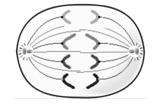
1) Draw a labelled diagram to show the metaphase stage of mitosis in an animal cell having ‘6’ chromosomes.
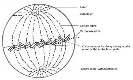
Ans: At metaphase in an animal cell with 6 chromosomes, the chromosomes (each appearing as an ‘X’ shape, indicating duplicated sister chromatids) would be lined up precisely along the metaphase plate (equatorial plane). Spindle fibers would extend from the centrosomes at opposite poles, attaching to the centromeres of each chromosome, ready for anaphase.
This diagram illustrates a metaphase stage, and adapts it for 6 chromosomes instead of the number shown. Ensure the labels include:
- Chromosomes aligned at the center
- Metaphase plate (or equatorial plane)
- Spindle fibers
- Centrosomes with centrioles (at the poles)
- Aster (radiating fibers around the centrosomes)
- Cytoplasm
2) The diagram below represents a stage during cell division. Study the same and then answer the question that follow:
a) Name the parts labelled 1, 2, and 3.
b) Identify the above stage and give a reason to support your answer.
c) Mention where in the body this type of cell division occurs.
d) Name the stage prior to this stage and draw a diagram to represent the same.
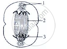
Ans: a) Labeled Parts: 1 – Aster, 2 – Spindle fibers, 3 – Chromosomes.
b) Stage Identification: Anaphase, as sister chromatids are visibly separating and moving towards opposing poles.
c) Location of Cell Division: Mitosis primarily occurs in somatic cells, facilitating processes like growth, tissue repair, and the replacement of old cells.
d) The stage prior to the one shown is Metaphase.
Diagram for Metaphase:
- A roughly circular cell outline.
- Clearly visible chromosomes aligned along the equatorial plate (the center of the cell).
- Each chromosome should consist of two sister chromatids.
- Spindle fibers extending from the poles of the cell and attaching to the centromeres of the chromosomes.
- Asters present at the poles (as it’s an animal cell, judging by the provided image).
3) The diagram given below represents a certain phenomenon which occurs during meiosis. Name and explain the phenomenon by using the terms – homologous chromosomes, chromatids, crossing over.
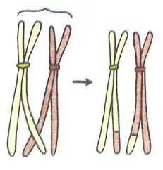
Ans: The diagram illustrates crossing over, a crucial phenomenon occurring during prophase I of meiosis.
During crossing over, homologous chromosomes (one from each parent, carrying genes for the same traits) pair up. Their non-sister chromatids (the arms of the paired chromosomes that are not identical copies of each other) physically intertwine and exchange segments of genetic material. This exchange results in recombinant chromatids, which contain a mix of genetic information from both parental chromosomes.
4) Given below is a diagram representing a stage during mitotic cell division in an animal cell examine it carefully and answer the questions which follow.
a) Identify the stage. Give one reason in support of your answer.
b) Name the cell organelle that forms the ‘aster’
c) Name the parts labelled 1, 2 and 3.
d) Name the stage that follows the one shown above, how is that stage identified?
e) Mention two differences between mitosis and meiosis with regards to: (i) The number of daughter cells produced. The chromosome number in the daughter cells.
Ans: a) The image you’ve provided clearly depicts the Anaphase stage of cell division, most likely mitosis.
The defining characteristic that allows for this identification is the separation of sister chromatids. In this stage, the centromeres, which previously held the sister chromatids together, divide. Consequently, each chromatid, now considered an individual chromosome, is pulled by the spindle fibers towards opposite poles of the cell. This movement is evident in the diagram, where the distinct V-shaped or J-shaped chromosomes are actively moving away from the equatorial plane and congregating at the two poles of the cell. The cell itself often begins to elongate during anaphase, foreshadowing the eventual division of the cytoplasm (cytokinesis).
b) The centrosome forms the aster.
c) 1: Centrosome/Centriole; 2: Spindle fibers; 3: Separating sister chromatids.
d) Telophase follows Anaphase, characterized by chromosomes reaching poles, decondensing, nuclear envelope reforming, and cytokinesis initiating.
e)Mitosis is a cell division process yielding two genetically identical, diploid daughter cells, crucial for growth, repair, and asexual reproduction, ensuring consistent genetic information. Meiosis, however, is a specialized division producing four genetically distinct, haploid daughter cells, essential for sexual reproduction.
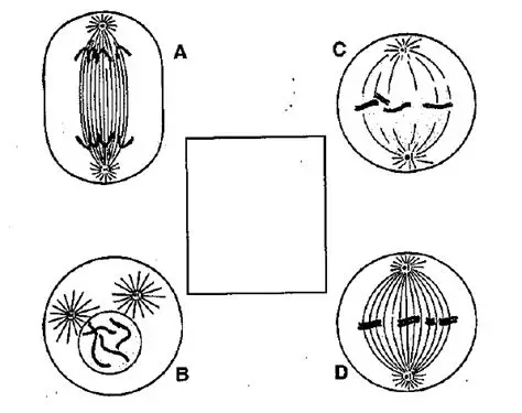
5) Given below are three diagrammatic sketches (A, B and C) of one and the same particular phase during mitotic type of cell division.
(a) Identify the phase
(b) What is the diploid number of chromosomes shown in them?
(c) Identify whether these are animal cells or plant cells?
(d) Which of these is/are shown in the correct direction? (i) Only A (ii) Only B (iii) Only A and C (iv) All the three
Ans: (a) Phase Identification: All three diagrams (A, B, and C) illustrate the anaphase of mitosis, characterized by the separation of sister chromatids moving to opposite poles.
(b) Diploid Chromosome Number: Each cell shows 6 chromosomes (3 moving to each pole) after chromatid separation, so the diploid number (2n) is 6.
(c) Cell Type Identification: A and B are animal cells, indicated by asters and no cell wall.
(d) Correct Direction: All three diagrams (A, B, and C) accurately depict the anaphase movement of chromosomes towards opposite poles. Thus, option (iv) All three are correct.
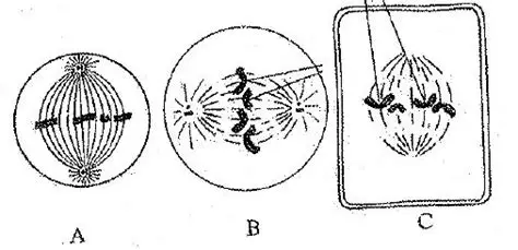
6) Shown below are four stages (A, B, C, D) (not in sequence) of a certain kind of cell division.
(a) Is it a plant cell or an animal cell? Give two reasons
(b) Is it undergoing mitosis or meiosis?
(c) What should be the correct sequence of these four stages among themselves?
(d) Name the stage that should precede the earliest of these stages
(e) Draw the stage names above inside the blank space provided.
Ans: (a) Animal Cell: The presence of asters and the absence of a cell wall in all diagrams indicate an animal cell.
(b) Mitosis: This is mitosis, as homologous chromosomes are not pairing or separating; instead, sister chromatids are aligning and separating.
(c) Correct Sequence: The correct sequence is B (Prophase), D (Metaphase), C (Anaphase), A (Telophase).
(d) Preceding Stage: Interphase precedes prophase.
(e) Drawing: (I cannot draw, but the stage preceding the earliest (Prophase, which is B) is Interphase. A diagram of interphase would show a nucleus with uncondensed chromatin and a visible nucleolus, with duplicated centrosomes nearby.)
