
")
The neural system is responsible for coordinating and integrating all the activities of the body’s organs. It consists of:
- Central Nervous System (CNS): Brain and spinal cord.
- Peripheral Nervous System (PNS): Cranial and spinal nerves.
Neurons are the functional units of the nervous system. They are specialized cells that transmit electrical signals called nerve impulses.
Nerve Impulse:
- Resting Potential: The difference in electrical charge across the resting neuron membrane.
- Action Potential: A wave of depolarization and repolarization that travels along the axon.
- Neurotransmitters: Chemical substances released at the synapse that allow the nerve impulse to be transmitted from one neuron to another.
Types of Neurons:
- Sensory Neurons: Carry sensory information from the body to the CNS.
- Interneurons: Connect sensory and motor neurons in the CNS.
- Motor Neurons: Carry commands from the CNS to muscles and glands.
Reflex Action:
- Involuntary responses to stimuli.
- Involve: Sensory neurons, interneurons, and motor neurons.
Central Nervous System:
- Brain: The control center of the body, responsible for complex functions like thought, memory, and emotions.
- Spinal Cord: Connects the brain to the rest of the body and carries sensory and motor information.
Peripheral Nervous System:
- Somatic Nervous System: Controls voluntary movements.
- Autonomic Nervous System: Controls involuntary functions like heart rate, blood pressure, and digestion.
Endocrine System:
- Works with the nervous system to coordinate body functions.
Key terms:
- Neurotransmitter: Acetylcholine, dopamine, serotonin, norepinephrine.
- Meninges: Protective layers surrounding the brain and spinal cord.
- Cerebrospinal Fluid: Fluid that protects and nourishes the brain and spinal cord.
Exercise
1. Briefly describe the structure of the Brain
Ans :
The human brain is a complex organ composed of billions of neurons and glial cells. It is divided into three main parts:
1. Cerebrum:
The largest part of the brain, responsible for higher-order functions such as thought, memory, language, and emotion.
Divided into four lobes: frontal, parietal, temporal, and occipital.
2. Cerebellum:
Located at the back of the brain, below the cerebrum.
Coordinates movement, balance, and posture.
3. Brainstem:
Connects the brain to the spinal cord.
Controls vital functions such as breathing, heart rate, and blood pressure.
Consists of the medulla oblongata, pons, and midbrain.
The brain is also divided into gray matter and white matter:
Gray matter: Contains the cell bodies of neurons and synapses.
White matter: Contains the axons of neurons, which are bundled together to form tracts that connect different parts of the brain.
2. Compare the following:
(a) Central neural system (CNS) and Peripheral neural system (PNS)
(b) Resting potential and action potential
Ans :
(a) Central neural system (CNS) and Peripheral neural system (PNS)
| Feature | Central Nervous System (CNS) | Peripheral Nervous System (PNS) |
| Components | Brain and spinal cord | Cranial nerves and spinal nerves |
| Function | Controls and coordinates all body functions | Relays information between the CNS and the body |
| Structure | Gray matter (cell bodies) and white matter (axons) | Sensory neurons, interneurons, and motor neurons |
| Role in Reflexes | Integrates sensory information and initiates motor responses | Contains sensory neurons that detect stimuli and motor neurons that carry out responses |
(b) Resting potential and action potential
| Feature | Resting Potential | Action Potential |
| Definition | The difference in electrical charge across the membrane of a neuron at rest | A brief, all-or-none electrical signal that travels down the axon of a neuron |
| Ion Distribution | Inside: High K+, low Na+ | Inside: High Na+, low K+ |
| Polarity | Negative inside, positive outside | Positive inside, negative outside |
| Purpose | Maintains the neuron’s readiness to fire an action potential | Transmits electrical signals along the neuron |
| Ion Channels | Leaky K+ channels | Voltage-gated Na+ and K+ channels |
3. Explain the following processes:
(a) Polarisation of the membrane of a nerve fibre
(b) Depolarisation of the membrane of a nerve fibre
(c) Transmission of a nerve impulse across a chemical synapse
Ans :
(a) Polarization of the Membrane of a Nerve Fiber
Polarization refers to the difference in electrical charge between the inside and outside of a neuron’s membrane. A neuron at rest is polarized, with the inside being negatively charged compared to the outside. This polarization is maintained by the selective permeability of the membrane to ions, primarily sodium (Na+) and potassium (K+).
- Sodium-Potassium Pump: This active transport mechanism pumps Na+ ions out of the cell and K+ ions into the cell, creating the concentration gradient that maintains the resting potential.
(b)
Depolarization occurs when the membrane potential becomes less negative or even positive. This happens when Na+ channels open, allowing Na+ ions to rush into the cell. This influx of positive ions neutralizes the negative charge inside the cell, leading to depolarization.
- Threshold Potential: If the depolarization reaches a certain threshold, it triggers an action potential.
(c) Transmission of a Nerve Impulse Across a Chemical Synapse
- Arrival of the Action Potential: The action potential reaches the presynaptic terminal of the neuron.
- Calcium Influx: Voltage-gated calcium channels open, allowing Ca2+ ions to enter the presynaptic terminal.
- Vesicle Fusion: The influx of Ca2+ triggers the fusion of synaptic vesicles containing neurotransmitters with the presynaptic membrane.
- Neurotransmitter Release: Neurotransmitters are released into the synaptic cleft, the space between the presynaptic and postsynaptic neurons.
- Binding to Receptors: Neurotransmitters bind to specific receptors on the postsynaptic membrane.
- Postsynaptic Potential: The binding of neurotransmitters can either depolarize (excitatory postsynaptic potential, EPSP) or hyperpolarize (inhibitory postsynaptic potential, IPSP) the postsynaptic neuron.
- Integration: The postsynaptic neuron integrates the effects of multiple EPSPs and IPSPs. If the net effect is depolarization and the threshold potential is reached, an action potential is generated in the postsynaptic neuron.
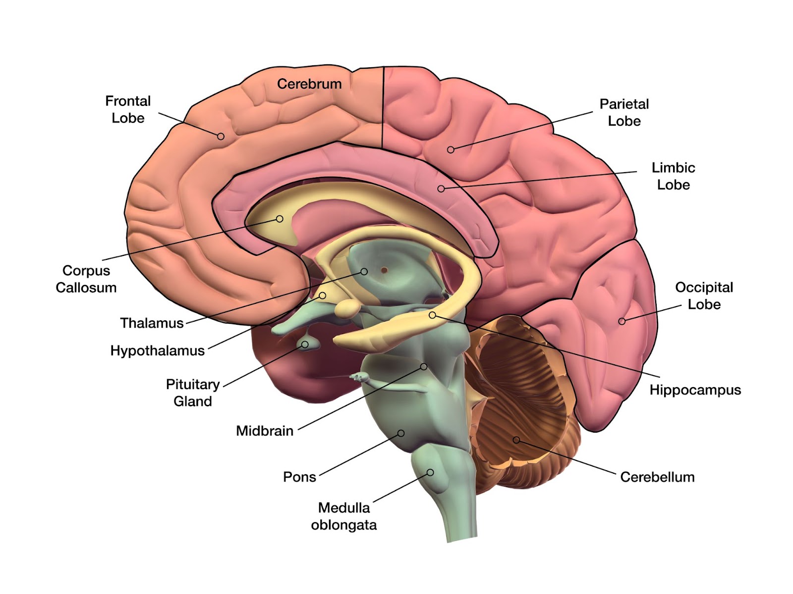
4. Draw labelled diagrams of the following: (a) Neuron (b) Brain
Ans :
(a) Neuron
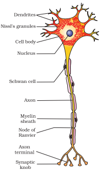
(b) Brain
5. Write short notes on the following:
(a) Neural coordination (b) Forebrain (c) Midbrain (d) Hindbrain (e) Synapse
Ans :
(a) Neural Coordination
- The nervous system coordinates and integrates various body functions.
- It consists of the central nervous system (CNS) and the peripheral nervous system (PNS).
- Neurons are the functional units of the nervous system.
- Nerve impulses are electrical signals transmitted along neurons.
- Synapses are the junctions between neurons where neurotransmitters facilitate communication.
(b) Forebrain
- The largest part of the brain, responsible for higher-order functions.
- Divided into lobes: frontal, parietal, temporal, and occipital.
- Involved in thought, memory, language, emotion, and sensory perception.
(c) Midbrain
- Located between the cerebrum and the pons.
- Involved in motor function, sensory perception, and arousal.
- Contains structures such as the substantia nigra, which plays a role in dopamine production and is involved in Parkinson’s disease.
(d) Hindbrain
- Located at the base of the brain.
- Consists of the medulla oblongata, pons, and cerebellum.
(e) Synapse
- Neurotransmitters are released at the synapse and bind to receptors on the postsynaptic neuron.
- Synaptic transmission can be excitatory or inhibitory, affecting the firing of the postsynaptic neuron.
6. Give a brief account of Mechanism of synaptic transmission
Ans :
Synaptic transmission is the process by which a nerve impulse is transmitted from one neuron (presynaptic neuron) to another neuron (postsynaptic neuron). It involves the release of chemical messengers called neurotransmitters.
Key steps in synaptic transmission:
- Action Potential Arrival: An action potential arrives at the presynaptic terminal, triggering the opening of voltage-gated calcium channels.
- Calcium Influx: Calcium ions (Ca2+) enter the presynaptic terminal.
- Vesicle Fusion: The influx of calcium ions causes synaptic vesicles containing neurotransmitters to fuse with the presynaptic membrane.
- Neurotransmitter Release: Neurotransmitters are released into the synaptic cleft, the space between the presynaptic and postsynaptic neurons.
- Binding to Receptors: Neurotransmitters bind to specific receptors on the postsynaptic membrane.
- Postsynaptic Potential: The binding of neurotransmitters can either depolarize (excitatory postsynaptic potential, EPSP) or hyperpolarize (inhibitory postsynaptic potential, IPSP) the postsynaptic neuron.
7. Explain the role of Na+ in the generation of action potential.
Ans :
Sodium (Na+) plays a crucial role in the generation of action potentials in neurons.
- Resting Potential: At rest, a neuron’s membrane is polarized, with the inside being negatively charged compared to the outside. This is primarily due to the higher concentration of potassium (K+) ions inside the cell and sodium (Na+) ions outside.
- Stimulus: When a neuron receives a stimulus (e.g., from another neuron or a sensory receptor), it causes voltage-gated sodium channels to open.
- Sodium Influx: Sodium ions rush into the cell, driven by the concentration gradient and the electrical potential difference.
- Depolarization: The influx of sodium ions causes the inside of the cell to become more positive, leading to depolarization.
- Positive Feedback Loop: Once the threshold is reached, more sodium channels open, leading to a rapid influx of sodium ions and a further increase in depolarization. This creates a positive feedback loop, ensuring that the action potential is all-or-none.
8. Differentiate between:
(a) Myelinated and non-myelinated axons
(b) Dendrites and axons
(c) Thalamus and Hypothalamus
(d) Cerebrum and Cerebellum
Ans :
(a) Myelinated and non-myelinated axons
| Feature | Myelinated Axon | Non-Myelinated Axon |
| Myelin Sheath | Covered with a fatty layer of myelin | Lacking a myelin sheath |
| Speed of Conduction | Faster | Slower |
| Saltatory Conduction | Yes (action potential jumps from node to node) | No (action potential spreads continuously) |
| Energy Efficiency | More energy-efficient | Less energy-efficient |
(b) Dendrites and axons
| Feature | Dendrites | Axons |
| Function | Receive signals from other neurons | Transmit signals to other neurons |
| Structure | Branched, tree-like | Long, unbranched |
| Direction of Signal | Towards the cell body | Away from the cell body |
| Myelin Sheath | May or may not be myelinated | Often myelinated |
(c) Thalamus and Hypothalamus
| Feature | Thalamus | Hypothalamus |
| Location | Above the brainstem | Below the thalamus |
| Function | Relays sensory information to the cerebral cortex, regulates sleep and wakefulness | Regulates body temperature, appetite, thirst, and hormone secretion |
| Role in Emotion | Plays a role in emotion and motivation | Plays a key role in the limbic system, regulating emotions |
(d) Cerebrum and Cerebellum
| Feature | Cerebrum | Cerebellum |
| Location | Largest part of the brain | Located below the cerebrum |
| Function | Higher-order functions (thought, memory, language, emotion) | Coordination of movement, balance, and posture |
| Structure | Divided into lobes (frontal, parietal, temporal, occipital) | Has a folded appearance with numerous gyri and sulci |
| Role in Learning and Memory | Plays a crucial role in learning and memory | Contributes to motor learning and memory |
9. Answer the following:
(a) Which part of the human brain is the most developed?
(b) Which part of our central neural system acts as a master clock?
Ans :
(a)It is responsible for higher-order functions such as thought, memory, language, and emotion.
(b) The hypothalamus acts as a master clock in our central nervous system. It regulates various biological rhythms, including the sleep-wake cycle, body temperature, and hormone secretion.
10. Distinguish between:
(a) afferent neurons and efferent neurons
(b) impulse conduction in a myelinated nerve fibre and unmyelinated nerve fibre
(c) cranial nerves and spinal nerves.
Ans :
(a) afferent neurons and efferent neurons
| Feature | Afferent Neurons | Efferent Neurons |
| Function | Carry sensory information from the body to the central nervous system (CNS) | Carry motor commands from the CNS to the body |
| Direction of Signal | Sensory (input) | Motor (output) |
| Examples | Sensory neurons in the skin, eyes, and ears | Motor neurons that control muscles and glands |
(b) impulse conduction in a myelinated nerve fibre and unmyelinated nerve fibre
| Feature | Myelinated Nerve Fiber | Unmyelinated Nerve Fiber |
| Myelin Sheath | Covered with a fatty myelin sheath | Lack a myelin sheath |
| Speed of Conduction | Faster (saltatory conduction) | Slower (continuous conduction) |
| Energy Efficiency | More energy-efficient | Less energy-efficient |
| Nodes of Ranvier | Have gaps in the myelin sheath called nodes of Ranvier | Lack nodes of Ranvier |
(c) cranial nerves and spinal nerves.
| Feature | Cranial Nerves | Spinal Nerves |
| Origin | Brain | Spinal cord |
| Number | 12 pairs | 31 pairs |
| Function | Control sensory and motor functions in the head and neck | Control sensory and motor functions in the body |
| Examples | Optic nerve, vagus nerve | Cervical nerves, thoracic nerves, lumbar nerves, sacral nerves, and coccygeal nerves |
